. 2009 Sep;28(7):416-24.
doi: 10.1016/j.matbio.2009.07.001. Epub 2009 Jul 25.

Characterization and regulation of ADAMTS-16

Affiliations

Characterization and regulation of ADAMTS-16

Alison K Surridge et al. Matrix Biol. 2009 Sep.

Abstract

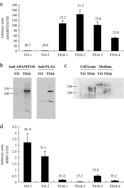
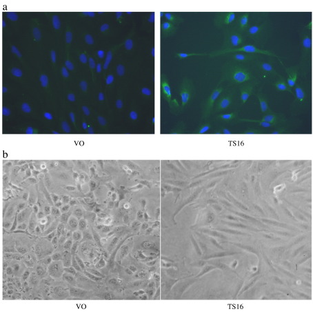
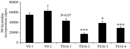
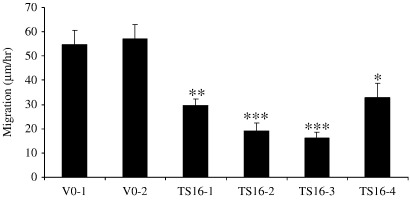
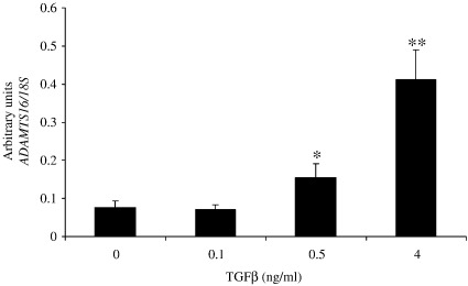
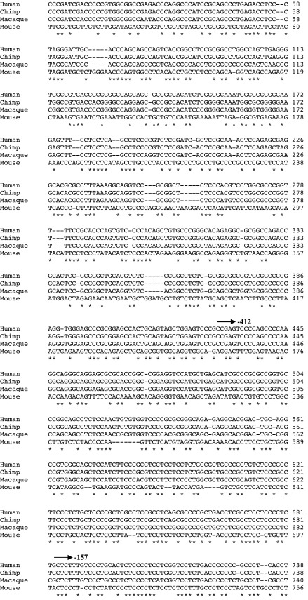
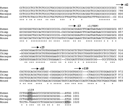
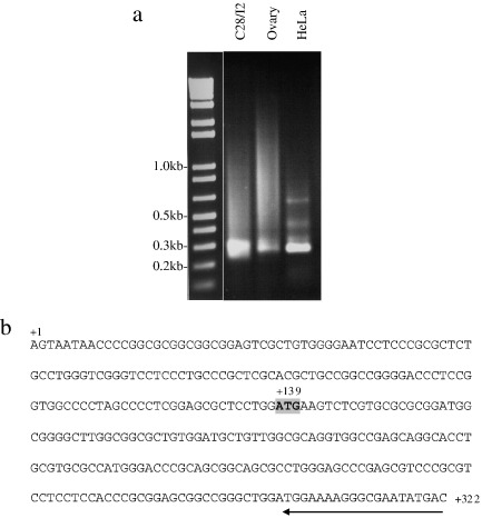
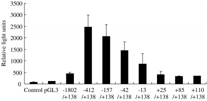
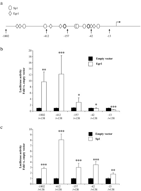
The ADAMTS (a disintegrin and metalloproteinase domain with thrombospondin motifs) family includes 19 secreted proteinases in man. ADAMTS16 is a recently cloned gene expressed at high levels in fetal lung and kidney and adult brain and ovary. The ADAMTS-16 protein currently has no known function. ADAMTS16 is also expressed in human cartilage and synovium where its expression is increased in tissues from osteoarthritis patients compared to normal tissues. In this study, we ascertained that the full length ADAMTS16 mRNA was expressed in chondrocytes and cloned the appropriate cDNA. Stable over-expression of ADAMTS16 in chondrosarcoma cells led to a decrease in cell proliferation and migration, though not adhesion, as well as a decrease in the expression of matrix metalloproteinase-13 (MMP13). The transcription start point of the human ADAMTS16 gene was experimentally identified as 138 bp upstream of the translation start ATG and the basal promoter was mapped out to -1802 bp. Overexpression of Egr1 induced ADAMTS16 promoter constructs of -157/+138 or longer whilst Sp1 induced all ADAMTS16 promoter constructs. Transforming growth factor beta (TGFbeta) stimulated expression of endogenous ADAMTS16 gene expression in chondrocyte cell lines.


Figures

Fig. 1
Fig. 1
Stable over-expression of ADAMTS16 in SW1353 cells. SW1353 cells were stably transfected with either vector only (VO) or ADAMTS16 (TS16) expression constructs. Cells were harvested and total RNA isolated and subjected to qRT-PCR for expression of (a) ADAMTS16 or (d) MMP13. Data are normalized to 18 S and expressed as mean ± s.e.m. Mean threshold cycle (Ct) is given above each bar. Extracellular matrix (b) or cell lysate and conditioned medium (c) were harvested (VO vs. TS16) and subjected to western blot analysis using anti-ADAMTS-16 and/or anti-FLAG primary antibodies.
Fig. 2
Fig. 2
Effect of ADAMTS-16 on cell phenotype. SW1353 cells were stably transfected with either vector only (VO) or ADAMTS16 (TS16) expression constructs. (a) Cells were fixed in 4% paraformaldehyde and stained with a 1:5000 dilution of mouse anti-FLAG antibody followed by FITC-linked second antibody; nuclei were counterstained with DAPI. (b) Cells were visualised under phase contrast 72 h after plating on plastic (×10 objective).
Fig. 3
Fig. 3
Proliferation of SW1353 cells stably expressing ADAMTS16. SW1353 cells were stably transfected with either vector only (VO) or ADAMTS16 (TS16) expression constructs. Cells were allowed to adhere overnight in DMEM/10% FCS then treated with 3H-thymidine in DMEM/0.5% FCS for 6 h followed by an overnight cold chase. Cell lysates were harvested and counted for tritium. Data are expressed as mean ± s.e.m., ⁎p < 0.05; ⁎⁎p < 0.01, ⁎⁎⁎p<0.001 comparing TS16 with either VO-1 or VO-2.
Fig. 4
Fig. 4
Cell migration of SW1353 cells stably expressing ADAMTS16. SW1353 cells were stably transfected with either vector only (VO) or ADAMTS16 (TS16) expression constructs. Eight hours after plating in DMEM/10% FCS, cells were subjected to time-lapse microscopy at 15 min intervals for 13 h. Distance moved per cell was averaged for 10 cells. Data are expressed as mean ± s.e.m., ⁎p < 0.05; ⁎⁎p < 0.01, ⁎⁎⁎p<0.001 comparing TS16 with VO-1 or VO-2.
Fig. 5
Fig. 5
Induction of ADAMTS16 expression by TGFβ. Confluent C28/I2 cells were serum starved for 24 h prior to addition of TGFβ for 6 h followed by harvest of RNA and qRT-PCR for ADAMTS16. Data are expressed as mean ± s.e.m., ⁎p < 0.05; ⁎⁎p < 0.01.
Fig. 6
Fig. 6
Sequence alignment for ADAMTS16 putative promoter region. Sequence alignment of 1000 bp upstream of ADAMTS16 ATG from human, macaque, chimp and mouse (www.ensembl.org). ⁎Indicates conserved regions. Translation start codon is shaded in grey with transcription start point (TSP) also marked. The 5′ positions of the deletion set of promoter–reporter constructs are also shown (arrows).
Fig. 6
Fig. 6
Sequence alignment for ADAMTS16 putative promoter region. Sequence alignment of 1000 bp upstream of ADAMTS16 ATG from human, macaque, chimp and mouse (www.ensembl.org). ⁎Indicates conserved regions. Translation start codon is shaded in grey with transcription start point (TSP) also marked. The 5′ positions of the deletion set of promoter–reporter constructs are also shown (arrows).
Fig. 7
Fig. 7
Identification of transcription start point in ADAMTS16 gene. 5′ RNA ligase-mediated (RLM) RACE was undertaken using C28/I2, human ovary or HeLa cell cDNA templates. (a) An agarose gel of the PCR products is shown; (b) following sequencing of RACE products, the transcription start point is marked as +1, translation start codon is shaded grey and position of RACE primer is shown (arrow).
Fig. 8
Fig. 8
Promoter activity in the ADAMTS16 gene. SW1353 cells were transiently transfected with promoter–reporter fragments in pGL3-basic as shown. Luciferase activity (measured as relative light units) of untransfected cells (control) and transient transfections of empty vector (pGL3) and eight deletion constructs ranging from − 1802/+138 to +110/+138 are plotted as mean ± s.e.m.
Fig. 9
Fig. 9
Activation of the ADAMTS16 promoter by Egr1 and Sp1. SW1353 cells were transiently transfected with promoter–reporter fragments in pGL3-basic as shown and co-transfected with expression constructs for Egr1, Sp1 or empty vector. Luciferase activity is plotted as fold compared to empty vector, mean ± s.e.m.; ⁎p < 0.05; ⁎⁎p < 0.01; ⁎⁎⁎p < 0.001. (a) schematic representation of Egr1 and Sp1 consensus sequences in the ADAMTS16 promoter; (b) induction of promoter by Egr1; (c) induction of promoter by Sp1.

References

    1. Blaney Davidson E.N., van der Kraan P.M., van den Berg W.B. TGF-beta and osteoarthritis. Osteoarthr. Cartil. 2007;15:597–604. - PubMed
    1. Cal S., Obaya A.J., Llamazares M., Garabaya C., Quesada V., Lopez-Otin C. Cloning, expression analysis, and structural characterization of seven novel human ADAMTSs, a family of metalloproteinases with disintegrin and thrombospondin-1 domains. Gene. 2002;283:49–62. - PubMed
    1. Chen S.J., Ning H., Ishida W., Sodin-Semrl S., Takagawa S., Mori Y., Varga J. The early-immediate gene EGR-1 is induced by transforming growth factor-beta and mediates stimulation of collagen gene expression. J. Biol. Chem. 2006;281:21183–21197. - PubMed
    1. Clark I.M., Parker A.E. Metalloproteinases: their role in arthritis and potential as therapeutic targets. Expert Opin. Ther. Targets. 2003;7:19–34. - PubMed
    1. Colige A., Sieron A.L., Li S.W., Schwarze U., Petty E., Wertelecki W., Wilcox W., Krakow D., Cohn D.H., Reardon W., Byers P.H., Lapiere C.M., Prockop D.J., Nusgens B.V. Human Ehlers–Danlos syndrome type VII C and bovine dermatosparaxis are caused by mutations in the procollagen I N-proteinase gene. Am. J. Hum. Genet. 1999;65:308–317. - PMC - PubMed

Publication types

LinkOut - more resources