. 2009 Oct;203(1):87-97.
doi: 10.1677/JOE-09-0065. Epub 2009 Jul 27.

Regulation of GNRH production by estrogen and bone morphogenetic proteins in GT1-7 hypothalamic cells

Affiliations

Regulation of GNRH production by estrogen and bone morphogenetic proteins in GT1-7 hypothalamic cells

Hiroyuki Otani et al. J Endocrinol. 2009 Oct.

Abstract

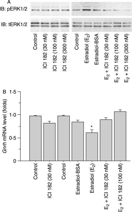
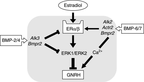
Recent studies have shown that bone morphogenetic proteins (BMPs) are important regulators in the pituitary-gonadal endocrine axis. We here investigated the effects of BMPs on GNRH production controlled by estrogen using murine GT1-7 hypothalamic neuron cells. GT1-7 cells expressed estrogen receptor alpha (ERalpha; ESR1 as listed in MGI Database), ERbeta (ESR2 as listed in MGI Database), BMP receptors, SMADs, and a binding protein follistatin. Treatment with BMP2 and BMP4 had no effect on Gnrh mRNA expression; however, BMP6 and BMP7 significantly increased Gnrh mRNA expression as well as GnRH production by GT1-7 cells. Notably, the reduction of Gnrh expression caused by estradiol (E(2)) was restored by cotreatment with BMP2 and BMP4, whereas it was not affected by BMP6 or BMP7. E(2) activated extracellular signal-regulated kinase (ERK) 1/2 and stress-activated protein kinase/c-Jun NH(2)-terminal kinase (SAPK/JNK) signaling but did not activate p38-mitogen-activated protein kinase (MAPK) signaling in GT1-7 cells. Inhibition of ERK1/ERK2 reversed the inhibitory effect of estrogen on Gnrh expression, whereas SAPK/JNK inhibition did not affect the E(2) actions. Expression levels of Eralpha and Erbeta were reduced by BMP2 and BMP4, but were increased by BMP6 and BMP7. Treatment with an ER antagonist inhibited the E(2) effects on Gnrh suppression including reduction of E(2)-induced ERK phosphorylation, suggesting the involvement of genomic ER actions in Gnrh suppression. BMP2 and BMP4 also suppressed estrogen-induced phosphorylation of ERK1/ERK2 and SAPK/JNK signaling, suggesting that BMP2 and BMP4 downregulate estrogen effects by attenuating ER-MAPK signaling. Considering that BMP6 and BMP7 increased the expression of alpha1E-subunit of R-type calcium channel (Cacna1e), which is critical for GNRH secretion, it is possible that BMP6 and BMP7 directly stimulate GNRH release by GT1-7 cells. Collectively, a newly uncovered interaction of BMPs and ER may be involved in controlling hypothalamic GNRH production and secretion via an autocrine/paracrine mechanism.


Figures

Figure 1
Figure 1
Expression of BMP system and estrogen receptor (ER) in GT1-7 cells. (A) Total cellular RNAs were extracted from GT1-7 cells and quantified by measuring the absorbance of the sample at 260 nm. The expression of mRNAs encoding Alk-2, -3, -4, -6, Bmpr2, Actr2a, Actr2b, Fst, Smad1–8, Gnrh, Erα, Erβ, and a housekeeping gene Rpl19 was examined by RT-PCR analysis in GT1-7 cells compared with a control sample extracted from mouse ovarian tissues. Aliquots of PCR products were electrophoresed on 1·5% agarose gel, visualized by ethidium bromide staining, and shown as representative of those obtained from three independent experiments. MM indicates molecular weight marker. (B) GT1-7 cells were precultured for 24 h and stimulated with BMP-2 and -4 (100 ng/ml) for 1 h. Immunofluorescence (IF) studies were performed using anti-phospho-SMAD1, 5, 8 (pSMAD1, 5, 8) antibody on BMP-treated cells. DAPI indicates counterstaining with 4′,6′-diaminodino-2-phenylindole. Bars indicate 20 μm in size.
Figure 2
Figure 2
Effects of BMPs on Gnrh transcription and Gnrh mRNA expression in GT1-7 cells. (A) GT1-7 cells (1×10⁵ cells/ml) were transiently transfected with Gnrh-luc reporter plasmid (500 ng) and pCMV-β-gal. After 24-h treatment with BMP-2, -4, -6, and -7 (100 ng/ml), cells were lysed and the luciferase activity was measured. The data were analyzed as the ratio of luciferase to β-galactosidase (β-gal) activity. Results are shown as mean±s.e.m. of data from at least three separate experiments, each performed with triplicate samples. **P<0·01 versus control groups. (B) GT1-7 cells (2×10⁵ cells/ml) were treated with BMP-2, -4, -6, and -7 (10 and 100 ng/ml) in serum-free conditions for 24 h. Total cellular RNA was extracted and Gnrh mRNA levels were examined by quantitative real-time RT-PCR. The expression levels of target genes were standardized by Rpl19 level in each sample. Results are shown as mean±s.e.m. of data from at least three separate experiments, each performed with triplicate samples. *P<0·05 versus control groups.
Figure 3
Figure 3
Effects of BMPs and estradiol on Gnrh transcription in GT1-7 cells. (A) GT1-7 cells (1×10⁵ cells/ml) were transiently transfected with Gnrh-luc reporter plasmid (500 ng) and pCMV-β-gal. After 24-h treatment with estradiol (1–100 nM), cells were lysed and the luciferase activity was measured. The data were analyzed as the ratio of luciferase to β-galactosidase (β-gal) activity. Results are shown as mean±s.e.m. of data from at least three separate experiments, each performed with triplicate samples. *P<0·05 versus control groups. (B) Cells (2×10⁵ cells/ml) were treated with BMP-2, -4, -6, and -7 (100 ng/ml) in the presence or absence of estradiol (100 nM) in serum-free conditions for 24 h. Total cellular RNA was extracted and Gnrh mRNA levels were examined by real-time RT-PCR. The expression levels of target genes were standardized by Rpl19 level in each sample. Results are shown as mean±s.e.m. of data from at least three separate experiments, each performed with triplicate samples. *P<0·05 versus control groups or between the indicated groups; NS, not significant.
Figure 4
Figure 4
Effects of BMPs on GNRH release by GT1-7 cells. (A) GT1-7 cells (2×10⁵ cells/ml) were treated with indicated concentrations of estradiol, BMP-2, -4, -6, and -7 in serum-free conditions. After 24-h culture, the culture media was collected, and GNRH concentrations (pg/ml) were determined by RIA. Results show the mean±s.e.m. of data performed with triplicate treatments; *P<0·05 versus control groups. (B) GT1-7 cells were treated with indicated concentrations of estradiol, BMP-2, -4, -6, and -7 in serum-free conditions for 24 h. Total cellular RNA was extracted and mRNA levels of Cacna1e were examined by real-time RT-PCR. The expression levels of target genes were standardized by Rpl19 level in each sample. Results are shown as mean±s.e.m. of data from at least three separate experiments, each performed with triplicate samples. *P<0·05 versus control groups.
Figure 5
Figure 5
Effects of BMPs and estradiol on GT1-7 cell proliferation. GT1-7 cells (1×10⁵ cells/ml) were treated with indicated concentrations of estradiol, BMP-2, -4, -6, -7, and PDGF-BB for 24 h in serum-free condition, and then thymidine uptake assay was performed. Results are shown as mean±s.e.m. of data from at least three separate experiments, each performed with triplicate samples. **P<0·01 versus control groups.
Figure 6
Figure 6
Effects of BMPs and estradiol on SMAD/MAPK signaling in GT1-7 cells. (A) Cells (1×10⁵ cells/ml) were precultured for 24 h in serum-free condition and stimulated with BMP-2, -4, -6, -7, and activin A (100 ng/ml) in the absence or presence of estradiol (100 nM). After 60-min culture, cells were lysed and subjected to SDS-PAGE/immunoblotting (IB) analysis using anti-phospho-SMAD1, 5, 8 (pSMAD1, 5, 8) and anti-SMAD5, anti-phospho-ERK1/2 (pERK1/2) and anti-total-ERK1/2 (tERK1/2), anti-phospho-SAPK/JNK (pSAPK/JNK) and anti-total-SAPK/JNK (tSAPK/JNK), anti-phospho-p38 (pp38) and anti-total-p38 (tp38), and anti-actin antibodies. The results shown are representative of those obtained from three independent experiments. (B) The relative integrated density of each protein band of Fig. 6A was digitized by NIH image J 1.34s. Results are shown as mean±s.e.m. of data from at least three separate experiments, each performed with triplicate samples. **P<0·01 and *P<0·05 versus control or basal levels.
Figure 7
Figure 7
Effects of MAPK inhibition on Gnrh regulation by GT1-7 cells. (A) GT1-7 cells (1×10⁵ cells/ml) were precultured for 24 h and then treated with MAPK inhibitors including U0126, SB203580, and SP600125 (3–10 μM) in combination with estradiol (E2; 100 nM). After 60-min culture, cells were lysed and subjected to SDS-PAGE/immunoblotting (IB) analysis using anti-phospho-ERK1/2 (pERK1/2), anti-total-ERK1/2 (tERK1/2), anti-phospho-SAPK/JNK (pSAPK/JNK), and anti-total SAPK/JNK (tSAPK/JNK) antibodies. The results shown are representative of those obtained from three independent experiments. (B) GT1-7 cells (2×10⁵ cells/ml) were treated with U0126 (3 μM) and SP600125 (10 μM) in the absence or presence of estradiol (100 nM) in serum-free conditions for 24 h. Total cellular RNA was extracted and Gnrh mRNA levels were examined by real-time RT-PCR. The expression levels of target genes were standardized by Rpl19 level in each sample. Results are shown as mean±s.e.m. of data from at least three separate experiments, each performed with triplicate samples. *P<0·05 versus control groups.
Figure 8
Figure 8
Effects of estrogen receptor (ER) inhibition on Gnrh regulation by GT1-7 cells. (A) GT1-7 cells (1×10⁵ cells/ml) were precultured for 24 h and then treated with ICI 182 780 (30–300 nM), estradiol (E2; 100 nM), and estradiol-BSA (100 nM). After 60-min culture, cells were lysed and subjected to SDS-PAGE/immunoblotting (IB) analysis using anti-phospho-ERK1/2 (pERK1/2) and anti-total-ERK1/2 (tERK1/2) antibodies. The results shown are representative of those obtained from three independent experiments. (B) GT1-7 cells (2×10⁵ cells/ml) were treated with ICI 182 780 (30–100 nM), estradiol (E2; 100 nM), and estradiol-BSA (100 nM) in serum-free conditions for 24 h. Total cellular RNA was extracted and Gnrh mRNA levels were examined by real-time RT-PCR. The expression levels of target genes were standardized by Rpl19 level in each sample. Results are shown as mean±s.e.m. of data from at least three separate experiments, each performed with triplicate samples. *P<0·05 versus control groups.
Figure 9
Figure 9
Effects of BMPs and estradiol on estrogen receptor (Er) expression in GT1-7 cells. (A and B) GT1-7 cells were treated with BMP-2, -4, -6, and -7 (10 and 100 ng/ml) and estradiol (100 nM) in serum-free conditions for 24 h. Total cellular RNA was extracted and Erα and Erβ mRNA levels were examined by real-time RT-PCR. The expression levels of target genes were standardized by Rpl19 level in each sample. Results are shown as mean±s.e.m. of data from at least three separate experiments, each performed with triplicate samples. *P<0·05 versus control groups. (C) Cells (1×10⁵ cells/ml) were treated with BMP-2, -4, -6, and -7 (100 ng/ml) in serum-free condition. After 24-h culture, cells were lysed and subjected to SDS-PAGE/immunoblotting (IB) analysis using anti-ERα, anti-ERβ, and anti-actin antibodies. The results shown are representative of those obtained from three independent experiments.
Figure 10
Figure 10
A possible mechanism by which BMPs and estrogen regulate GNRH production by GT1-7 cells. Estradiol suppresses GNRH production by inhibiting ERK1/ERK2 pathway via ER. In this feedback system between estradiol and GNRH production, BMP2 and BMP4 downregulate estrogen effects by attenuating estrogen-ERK signaling and ERα and ERβ expression. In contrast, BMP6 and BMP7 directly stimulate Gnrh transcription, GNRH production, and GNRH release via increased expression of α1E-subunit of R-type calcium channel, which is critical for GNRH secretion. A new interaction of BMPs and ER action may be involved in controlling hypothalamic GNRH production and secretion in an autocrine/paracrine mechanism.

Similar articles

Cited by

References

    1. Avola R, Spina-Purrello V, Gallo F, Morale MC, Marletta N, Costa A, Tirolo C, Testa N, Reale S, Marchetti B. Immortalized hypothalamic luteinizing hormone-releasing hormone (LHRH) neurons induce a functional switch in the growth factor responsiveness of astroglia: involvement of basic fibroblast growth factor. International Journal of Developmental Neuroscience. 2000;18:743–763. - PubMed
    1. Bowe J, Li XF, Sugden D, Katzenellenbogen JA, Katzenellenbogen BS, O'Byrne KT. The effects of the phytoestrogen, coumestrol, on gonadotropin-releasing hormone (GnRH) mRNA expression in GT1-7 GnRH neurones. Journal of Neuroendocrinology. 2003;15:105–108. - PubMed
    1. Buchanan CD, Mahesh VB, Brann DW. Estrogen-astrocyte-luteinizing hormone-releasing hormone signaling: a role for transforming growth factor-beta(1). Biology of Reproduction. 2000;62:1710–1721. - PubMed
    1. Chappell PE, White RS, Mellon PL. Circadian gene expression regulates pulsatile gonadotropin-releasing hormone (GnRH) secretory patterns in the hypothalamic GnRH-secreting GT1-7 cell line. Journal of Neuroscience. 2003;23:11202–11213. - PMC - PubMed
    1. Charles AC, Hales TG. Mechanisms of spontaneous calcium oscillations and action potentials in immortalized hypothalamic (GT1-7) neurons. Journal of Neurophysiology. 1995;73:56–64. - PubMed

Publication types

MeSH terms