Review
. 2009 Sep-Oct;100(5):624-36.
doi: 10.1093/jhered/esp063. Epub 2009 Jul 27.

A surrogate approach to study the evolution of noncoding DNA elements that organize eukaryotic genomes

Affiliations
Review

A surrogate approach to study the evolution of noncoding DNA elements that organize eukaryotic genomes

Danielle Vermaak et al. J Hered. 2009 Sep-Oct.

Abstract

Comparative genomics provides a facile way to address issues of evolutionary constraint acting on different elements of the genome. However, several important DNA elements have not reaped the benefits of this new approach. Some have proved intractable to current day sequencing technology. These include centromeric and heterochromatic DNA, which are essential for chromosome segregation as well as gene regulation, but the highly repetitive nature of the DNA sequences in these regions make them difficult to assemble into longer contigs. Other sequences, like dosage compensation X chromosomal sites, origins of DNA replication, or heterochromatic sequences that encode piwi-associated RNAs, have proved difficult to study because they do not have recognizable DNA features that allow them to be described functionally or computationally. We have employed an alternate approach to the direct study of these DNA elements. By using proteins that specifically bind these noncoding DNAs as surrogates, we can indirectly assay the evolutionary constraints acting on these important DNA elements. We review the impact that such "surrogate strategies" have had on our understanding of the evolutionary constraints shaping centromeres, origins of DNA replication, and dosage compensation X chromosomal sites. These have begun to reveal that in contrast to the view that such structural DNA elements are either highly constrained (under purifying selection) or free to drift (under neutral evolution), some of them may instead be shaped by adaptive evolution and genetic conflicts (these are not mutually exclusive). These insights also help to explain why the same elements (e.g., centromeres and replication origins), which are so complex in some eukaryotic genomes, can be simple and well defined in other where similar conflicts do not exist.


Figures

Figure 1
Figure 1
A “surrogate” strategy to study noncoding DNA. In the schematic figure, the evolution of the underlying noncoding DNA is difficult to decipher because the sequence of the DNA is either unknown or difficult to describe. However, by studying the pattern of evolution acting specifically on the protein–DNA interfaces (hashed region) of proteins that specifically bind these DNAs, we can infer the types of changes that must have taken place on the underlying DNA, even in the absence of knowledge of what those DNA sequences are. Thus, the DNA-binding proteins serve as a surrogate allowing us to study the evolutionary constraints acting on important architectural noncoding DNAs that are hard to study otherwise.
Figure 2
Figure 2
Surrogate strategy to study centromeric DNA. (A) “Surrogate” centromere proteins. Proteins like centromeric histones intimately associate with centromeric DNA sequence to directly identify the centromere. Heterochromatin proteins, on the other hand, indirectly define the centromere by establishing a boundary to the centromere. Evolutionary studies on either set of proteins provide novel hypotheses regarding the evolutionary dynamics and function of the underlying centromeric/pericentromeric DNA. (B) “Centromere-drive” model. Paired homologous chromosomes in metaphase I are illustrated. Centromeres (dark gray) are shown with an assembled kinetochore (stippled) bound to microtubules. In this example, the size of the kinetochore is dependent on the amount of centromeric sequence. In step 1, a novel centromeric satellite (white) expands and the new, larger centromere can drive in female meiosis by attracting more microtubules and gaining a favorable position in asymmetric female meiosis. This results in a selective advantage for this particular centromere, which will quickly fix in the population. In step 2, pairing of centromeres in male meiosis with unequal strengths results in unequal tension across the centromeres. Unequal tension can potentially increase the rate of nondisjunction in males, resulting in sterility. Selection acts on the DNA-binding affinity of proteins to restore parity across the centromeres and suppresses drive and sterility effects (shown here as restricting 1 centromere). Repeated bouts of drive and suppression will be observed as positive selection among centromere or heterochromatin proteins and fixation of new centromeric sequences (Henikoff and Malik 2002; Malik and Bayes 2006).
Figure 3
Figure 3
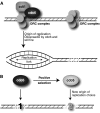
Surrogate strategy to study origins of DNA replication. (A) “Surrogate” DNA replication proteins. Several proteins mediate the accurate, coordinated firing of multiple origins of DNA replication in eukaryotic genomes. For simplicity, we are only showing the ORC complex proteins that define origins, of which only a subset that recruits Cdc6 and Cdt1 will be “licensed” to result in productive replication origins. (B) Altering the genomic landscape of DNA replication. Cdc6 has undergone positive selection in 2 separate lineages of Drosophila. This leads to the model where adaptive evolution has changed the DNA-binding specificity of Cdc6 and thereby the pattern of replication origins. One mechanism by which this could occur is through changes in Cdc6-binding affinity altering the probability of replication licensing at different DNA sequences (Speck et al. 2005; Speck and Stillman 2007).
Figure 4
Figure 4
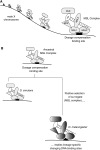
Surrogate strategy to study dosage compensation X chromosomal sites. (A) “Surrogate” dosage compensation proteins. The MSL complex binds to many sites on the male X chromosome, and this binding is critical for dosage compensation and viability of Drosophila males. MSL1 and MSL2 play a key role in targeting of the complex to the X chromosome. (B) Alterations of dosage compensation X chromosomal sites. The MSL complex is evolving under positive selection in the Drosophila melanogaster lineage, but not in the Drosophila simulans lineage (Levine et al. 2007; Rodriguez et al. 2007). Positive selection appears to be concentrated in domains of MSL1 and MSL2 that are critical for targeting to the X chromosome (Rodriguez et al. 2007). This suggests that DNA-binding sites are rapidly changing in a lineage-specific manner, a prediction that has recently been confirmed (Bachtrog 2008).

Similar articles

Cited by

References

    1. Ahmad K, Henikoff S. Histone H3 variants specify modes of chromatin assembly. Proc Natl Acad Sci USA. 2002;99(Suppl 4):16477–16484. - PMC - PubMed
    1. Akhtar A, Becker PB. Activation of transcription through histone H4 acetylation by MOF, an acetyltransferase essential for dosage compensation in Drosophila. Mol Cell. 2000;5:367–375. - PubMed
    1. Alekseyenko AA, Larschan E, Lai WR, Park PJ, Kuroda MI. High-resolution ChIP-chip analysis reveals that the Drosophila MSL complex selectively identifies active genes on the male X chromosome. Genes Dev. 2006;20:848–857. - PMC - PubMed
    1. Alekseyenko AA, Peng S, Larschan E, Gorchakov AA, Lee OK, Kharchenko P, McGrath SD, Wang CI, Mardis ER, Park PJ, et al. A sequence motif within chromatin entry sites directs MSL establishment on the Drosophila X chromosome. Cell. 2008;134:599–609. - PMC - PubMed
    1. Assis R, Kondrashov AS. Rapid repetitive element-mediated expansion of piRNA clusters in mammalian evolution. Proc Natl Acad Sci USA. 2009; 106:7079–7082. - PMC - PubMed

Publication types