

Dissection of the endogenous cellular pathways of PCSK9-induced low density lipoprotein receptor degradation: evidence for an intracellular route
- PMID: 19635789
- PMCID: PMC2781431
- DOI: 10.1074/jbc.M109.037085
Dissection of the endogenous cellular pathways of PCSK9-induced low density lipoprotein receptor degradation: evidence for an intracellular route
Abstract
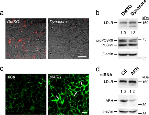
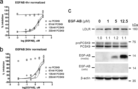
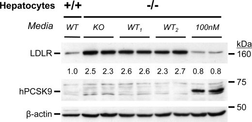
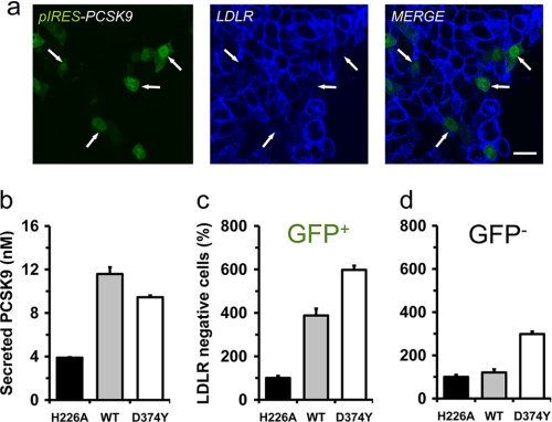
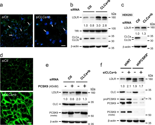
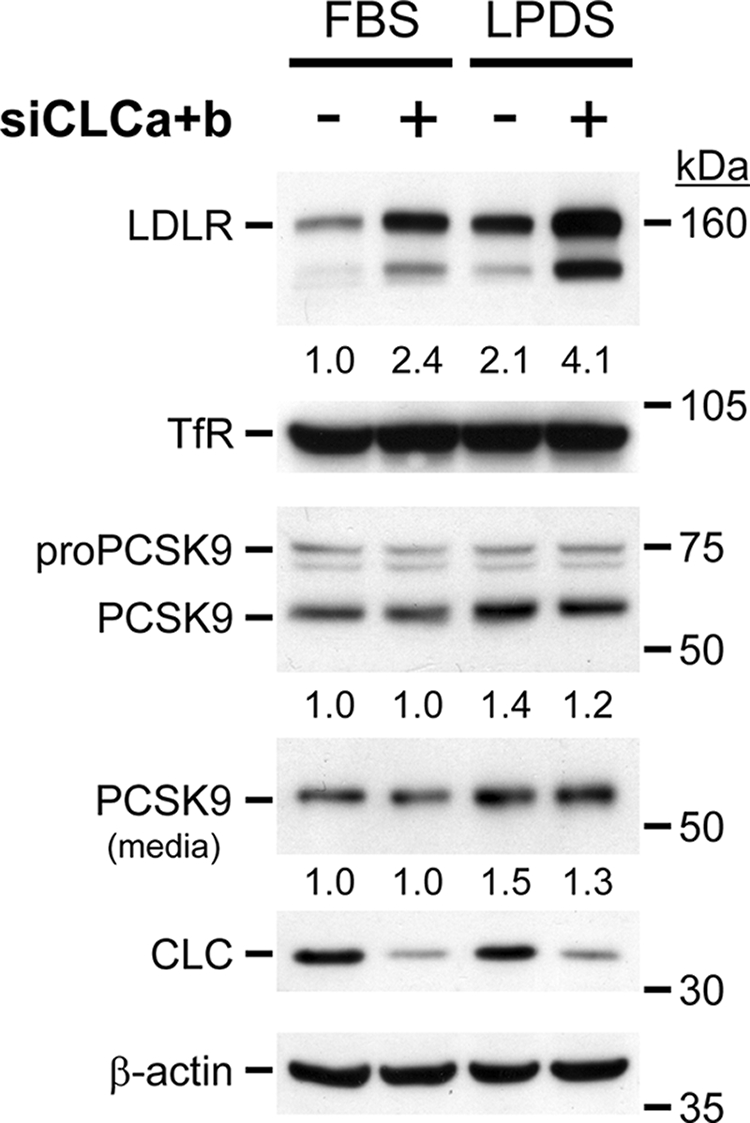
Elevated levels of plasma low density lipoprotein (LDL)-cholesterol, leading to familial hypercholesterolemia, are enhanced by mutations in at least three major genes, the LDL receptor (LDLR), its ligand apolipoprotein B, and the proprotein convertase PCSK9. Single point mutations in PCSK9 are associated with either hyper- or hypocholesterolemia. Accordingly, PCSK9 is an attractive target for treatment of dyslipidemia. PCSK9 binds the epidermal growth factor domain A (EGF-A) of the LDLR and directs it to endosomes/lysosomes for destruction. Although the mechanism by which PCSK9 regulates LDLR degradation is not fully resolved, it seems to involve both intracellular and extracellular pathways. Here, we show that clathrin light chain small interfering RNAs that block intracellular trafficking from the trans-Golgi network to lysosomes rapidly increased LDLR levels within HepG2 cells in a PCSK9-dependent fashion without affecting the ability of exogenous PCSK9 to enhance LDLR degradation. In contrast, blocking the extracellular LDLR endocytosis/degradation pathway by a 4-, 6-, or 24-h incubation of cells with Dynasore or an EGF-AB peptide or by knockdown of endogenous autosomal recessive hypercholesterolemia did not significantly affect LDLR levels. The present data from HepG2 cells and mouse primary hepatocytes favor a model whereby depending on the dose and/or incubation period, endogenous PCSK9 enhances the degradation of the LDLR both extra- and intracellularly. Therefore, targeting either pathway, or both, would be an effective method to reduce PCSK9 activity in the treatment of hypercholesterolemia and coronary heart disease.
Figures
Similar articles
-
The cellular trafficking of the secretory proprotein convertase PCSK9 and its dependence on the LDLR.Traffic. 2007 Jun;8(6):718-32. doi: 10.1111/j.1600-0854.2007.00562.x. Epub 2007 Apr 25. Traffic. 2007. PMID: 17461796
-
Effects of pH and low density lipoprotein (LDL) on PCSK9-dependent LDL receptor regulation.J Biol Chem. 2007 Jul 13;282(28):20502-12. doi: 10.1074/jbc.M701634200. Epub 2007 May 10. J Biol Chem. 2007. PMID: 17493938
-
The proprotein convertase PCSK9 induces the degradation of low density lipoprotein receptor (LDLR) and its closest family members VLDLR and ApoER2.J Biol Chem. 2008 Jan 25;283(4):2363-72. doi: 10.1074/jbc.M708098200. Epub 2007 Nov 26. J Biol Chem. 2008. PMID: 18039658
-
Sorting an LDL receptor with bound PCSK9 to intracellular degradation.Atherosclerosis. 2014 Nov;237(1):76-81. doi: 10.1016/j.atherosclerosis.2014.08.038. Epub 2014 Sep 2. Atherosclerosis. 2014. PMID: 25222343 Review.
-
Targeting the proprotein convertase subtilisin/kexin type 9 for the treatment of dyslipidemia and atherosclerosis.J Am Coll Cardiol. 2013 Oct 15;62(16):1401-8. doi: 10.1016/j.jacc.2013.07.056. Epub 2013 Aug 21. J Am Coll Cardiol. 2013. PMID: 23973703 Review.
Cited by
-
Proprotein Convertase Subtilisin/Kexin Type 9 (PCSK9) Single Domain Antibodies Are Potent Inhibitors of Low Density Lipoprotein Receptor Degradation.J Biol Chem. 2016 Aug 5;291(32):16659-71. doi: 10.1074/jbc.M116.717736. Epub 2016 Jun 8. J Biol Chem. 2016. PMID: 27284008 Free PMC article.
-
Monoclonal Antibodies for Lipid Management.Curr Atheroscler Rep. 2016 Jul;18(7):39. doi: 10.1007/s11883-016-0593-2. Curr Atheroscler Rep. 2016. PMID: 27221501 Review.
-
Molecular Regulation and Therapeutic Targeting of VLDL Production in Cardiometabolic Disease.Cell Mol Gastroenterol Hepatol. 2025;19(1):101409. doi: 10.1016/j.jcmgh.2024.101409. Epub 2024 Oct 12. Cell Mol Gastroenterol Hepatol. 2025. PMID: 39406347 Free PMC article. Review.
-
Lowering low-density lipoprotein cholesterol: from mechanisms to therapies.Life Metab. 2022 May 20;1(1):25-38. doi: 10.1093/lifemeta/loac004. eCollection 2022 Aug. Life Metab. 2022. PMID: 39872686 Free PMC article. Review.
-
PCSK9 promotes progression of anaplastic thyroid cancer through E-cadherin endocytosis.Cell Death Dis. 2025 May 6;16(1):362. doi: 10.1038/s41419-025-07690-1. Cell Death Dis. 2025. PMID: 40328788 Free PMC article.
References
-
- Lloyd-Jones D., Adams R., Carnethon M., De Simone G., Ferguson T. B., Flegal K., Ford E., Furie K., Go A., Greenlund K., Haase N., Hailpern S., Ho M., Howard V., Kissela B., Kittner S., Lackland D., Lisabeth L., Marelli A., McDermott M., Meigs J., Mozaffarian D., Nichol G., O'Donnell C., Roger V., Rosamond W., Sacco R., Sorlie P., Stafford R., Steinberger J., Thom T., Wasserthiel-Smoller S., Wong N., Wylie-Rosett J., Hong Y. (2009) Circulation 119, e21–e181 - PubMed
-
- Brown M. S., Goldstein J. L. (1986) Science 232, 34–47 - PubMed
-
- Varret M., Abifadel M., Rabès J. P., Boileau C. (2008) Clin. Genet. 73, 1–13 - PubMed
-
- Seidah N. G., Prat A. (2007) J. Mol. Med. 85, 685–696 - PubMed
Publication types
MeSH terms
Substances
LinkOut - more resources
Full Text Sources
Other Literature Sources
Miscellaneous
