Comparative Study
. 2009 Oct 23;284(43):29773-83.
doi: 10.1074/jbc.M109.032144. Epub 2009 Jul 27.

Structural basis for the mechanism of respiratory complex I

Affiliations
Comparative Study

Structural basis for the mechanism of respiratory complex I

John M Berrisford et al. J Biol Chem. .

Abstract

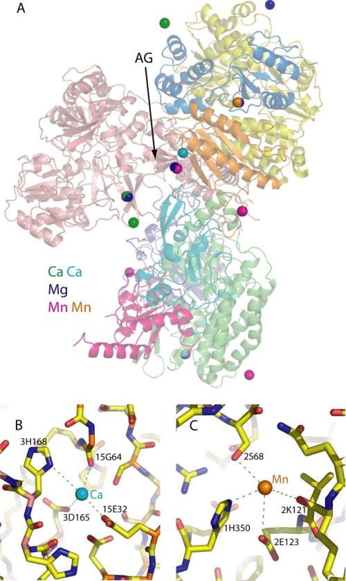
Complex I plays a central role in cellular energy production, coupling electron transfer between NADH and quinone to proton translocation. The mechanism of this highly efficient enzyme is currently unknown. Mitochondrial complex I is a major source of reactive oxygen species, which may be one of the causes of aging. Dysfunction of complex I is implicated in many human neurodegenerative diseases. We have determined several x-ray structures of the oxidized and reduced hydrophilic domain of complex I from Thermus thermophilus at up to 3.1 A resolution. The structures reveal the mode of interaction of complex I with NADH, explaining known kinetic data and providing implications for the mechanism of reactive oxygen species production at the flavin site of complex I. Bound metals were identified in the channel at the interface with the frataxin-like subunit Nqo15, indicating possible iron-binding sites. Conformational changes upon reduction of the complex involve adjustments in the nucleotide-binding pocket, as well as small but significant shifts of several alpha-helices at the interface with the membrane domain. These shifts are likely to be driven by the reduction of nearby iron-sulfur clusters N2 and N6a/b. Cluster N2 is the electron donor to quinone and is coordinated by unique motif involving two consecutive (tandem) cysteines. An unprecedented "on/off switch" (disconnection) of coordinating bonds between the tandem cysteines and this cluster was observed upon reduction. Comparison of the structures suggests a novel mechanism of coupling between electron transfer and proton translocation, combining conformational changes and protonation/deprotonation of tandem cysteines.


Figures

FIGURE 1.
FIGURE 1.
The NADH-binding site. A, the site viewed from the solvent-exposed side. FMN and residues involved in NADH binding are shown as sticks with carbon in yellow and NADH with carbon in salmon. Hydrogen bonds are shown as dotted lines in green, stacking interactions are in gray, and the hydride transfer path is in red. The van der Waal's contact between 1Glu97 and C4N of NADH is shown in gray. Prefixes before residue names indicate the subunit number. B, conformational changes in the nucleotide-binding site upon reduction by NADH. Superposed structures of the oxidized (O2, green) and NADH-reduced (RND, yellow) domain are shown. C, surface representation without NADH; FMN is shown as sticks with carbon in cyan. D, surface representation with bound NADH, colored salmon.
FIGURE 2.
FIGURE 2.
Conformational changes upon reduction by NADH. A and B, the structure of the NADH-reduced domain (RND) was superposed with that of oxidized domain (O2) and colored according to Cα deviations between the structures - blue to green to yellow as deviation increases. Higher deviations near the nucleotide-binding site (top) and at the interface with the membrane domain (bottom) are evident. NADH is shown as spheres in magenta. C, close-up of the interface with the membrane domain (structures RND and O4 were superposed). The approximate (expected) border of the membrane domain is shaded in blue. Helices H1 and H2 from Nqo6 subunit and the four-helix bundle from Nqo4 are indicated. Clusters N2 and N6b are shown as spheres. The arrows indicate movements of helix H1 and the four-helix bundle upon reduction.
FIGURE 3.
FIGURE 3.
Environment of cluster N2. A and B, oxidized domain (structure O2). Two copies of the molecule in the ASU are shown. C and D, NADH-reduced domain (structure RND). Two copies of the molecule in the ASU are shown. E, dithionite-reduced domain (structure RD). F, NADH-reduced domain exposed to air for ∼30 min (structure RA1). Cluster ligands and residues discussed in the text are shown as sticks. The backbones of subunits are colored green for Nqo4 and red for Nqo6. Electron density is from a σA-weighted 2Fo − Fc maps contoured at 1σ. The cluster is shown as spheres of 0.3 van der Waal's radius. The prefixes before the residue names indicate the subunit number.
FIGURE 4.
FIGURE 4.
Bound cations. A, an overview. The three structures (O4, O2, and RND) containing three different types of bound cations were superposed, and the cations are shown as spheres. Oxidized domain structure (O4) is shown in cartoon representation with subunit Nqo1 is in yellow, Nqo2 is in blue, Nqo3 is in salmon, Nqo4 is in green, Nqo5 is in violet, Nqo6 is in red, Nqo9 is in cyan, and Nqo15 is in orange. AG indicates the acidic groove. Ca2+ ions are shown in green except the tightly bound Ca2+ present in all structures, which is in cyan. Mg2+ is in blue, and Mn2+ is in magenta except the Mn2+ atom bound in the putative iron-binding channel, which is in orange. B, close-up of the tightly bound Ca2+ ion with interacting residues indicated. C, close-up of the Mn2+ ion bound in the putative iron-binding channel at the interface with frataxin-like subunit Nqo15. Residues interacting with the metal are indicated, including possible weak interaction with carbonyl of 2Lys121. The prefixes before the residue names indicate the subunit number.
FIGURE 5.
FIGURE 5.
A model of complex I mechanism, based on structures described here. Iron-sulfur clusters N2 and N6b are depicted as oxidized or reduced by empty or filled circles, respectively. C46 and C45 indicate the tandem cysteines from Nqo6 subunit, whereas Q/QH2 indicate quinone/quinol. One of possible proton acceptors from C45 is 4Tyr87 (Y-O) and from C46-6Glu49 (E-O). H-path indicates proton delivery pathway from the cytosol to tandem cysteines, such as that shown in supplemental Fig. 5, which may be the same for both cysteines (although other pathways are also possible). H1 and 4HB indicate helix H1 from Nqo6 and the four-helix bundle from Nqo4, respectively, and are shown on dark backgrounds when helices are shifted relative to oxidized state. The proposed catalytic cycle is described in detail in the text.

Similar articles

Cited by

References

    1. Walker J. E. (1992) Q. Rev. Biophys. 25, 253–324 - PubMed
    1. Yagi T., Matsuno-Yagi A. (2003) Biochemistry 42, 2266–2274 - PubMed
    1. Carroll J., Fearnley I. M., Skehel J. M., Shannon R. J., Hirst J., Walker J. E. (2006) J. Biol. Chem. 281, 32724–32727 - PubMed
    1. Sazanov L. A. (2007) Biochemistry 46, 2275–2288 - PubMed
    1. Schapira A. H. (1998) Biochim. Biophys. Acta 1364, 261–270 - PubMed

Publication types

MeSH terms