. 2009 Sep 15;587(Pt 18):4455-66.
doi: 10.1113/jphysiol.2009.173468. Epub 2009 Jul 27.

Electrical coupling synchronises spinal motoneuron activity during swimming in hatchling Xenopus tadpoles

Affiliations

Electrical coupling synchronises spinal motoneuron activity during swimming in hatchling Xenopus tadpoles

Hong-Yan Zhang et al. J Physiol. .

Abstract

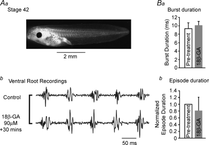
The role of electrical coupling between neurons in the swimming rhythm generator of Xenopus embryos has been studied using pharmacological blockade of gap junctions. A conspicuous effect of 18beta-glycyrrhetinic acid (18beta-GA) and carbenoxolone, which have been shown to block electrical coupling in this preparation, was to increase the duration of ventral root bursts throughout the spinal cord during swimming. The left-right coordination, the swimming frequency and the duration of swimming episodes were not affected by concentrations of 18beta-GA which significantly increased burst durations. However, the longitudinal coupling was affected such that 18beta-GA led to a significant correlation between rostrocaudal delays and cycle periods, which is usually only present in older larval animals. Patch clamp recordings from spinal motoneurons tested whether gap junction blockers affect the spike timing and/or firing pattern of motoneurons during fictive swimming. In the presence of 18beta-GA motoneurons continued to fire a single, but broader action potential in each cycle of swimming, and the timing of their spikes relative to the ventral root burst became more variable. 18beta-GA had no detectable effect on the resting membrane potential of motoneurons, but led to a significant increase in input resistance, consistent with the block of gap junctions. This effect did not result in increased firing during swimming, despite the fact that multiple spikes can occur in response to current injection. Applications of 18beta-GA at larval stage 42 had no discernible effect on locomotion. The results, which suggest that electrical coupling primarily functions to synchronize activity in synergistic motoneurons during embryo swimming, are discussed in the context of motor system development.


Figures

Figure 1
Figure 1. The effects of gap junction blockers on swimming bursts and the electrical coupling of hindbrain dINs of Xenopus embryos
Aa, Xenopus tadpole at stage 37/38, from Nieuwkoop & Faber (1956). Ab and c, effects of two gap junction blockers, 18β-GA (90 μm) and CBNX (100 μm), and d, a time control, on ventral root bursts during fictive swimming. B, the increase in ventral root burst durations in 18β-GA correlates temporally with the blocking of electrical coupling between hdINs. Ba, ventral root burst duration measured on the 10th cycle at the beginning of each swimming episode after applying 40 μm 18β-GA; grey: individual measurements from 6 preparations; black: mean durations ±s.d.; Bb, normalised average electrical coupling coefficients (same preparations as Ba) after 18β-GA applied at time zero (see Li et al. 2009). Bc, burst durations measured on the 10th cycle at the beginning of each swimming episode do not change with time under control conditions; grey: individual measurements from 5 preparations; black: average durations ±s.d. Bd, simultaneous whole-cell patch recordings of coupled dINs in control (left column) and after 18β-GA treatment (right column). Upper trace: one example of the 10th ventral root bursts from the beginning of swimming before and after 18β-GA blockade; lower traces: responses of two coupled dINs to hyperpolarizing current injected in either cell. Recording time points of control and block examples were indicated by left and right arrows in Ba, respectively.
Figure 2
Figure 2. Dose-dependent differentiation of 18β-GA effects on fictive swimming of Xenopus embryos
In each preparation (n= 7) the concentration was maintained at the specified level (0, 15, 30, 90 μm) for 30 min, and then increased to the next higher concentration. Significant effects on ventral root burst duration (A) occur at lower concentrations (30 μm) than effects on swimming episode duration (B; 90 μm) and swimming cycle period (C; 90 μm). *P < 0.05; **P < 0.01.
Figure 3
Figure 3. Distributed effects of a low concentration of 18β-GA (30 μm) on fictive swimming
A, example of ventral root recordings in pre-treatment control (a) and 30 min after 18β-GA application (b). L4 and L11: recording from the fourth and eleventh intermyotomal cleft on left side, respectively; R8: recording from the eighth cleft on right side. B–D, white bars = time 0 (pre-treatment); shaded bars =+30 min (either treatment or control). B, control data (n= 5) and effects of 18β-GA (n= 7) and CBNX (n= 6) on swimming burst durations. a and b: rostral (L4); c and d caudal (L11) burst durations. Asterisks indicate significant differences between time 0 and 30 min. C, 30 μm 18β-GA did not affect left–right coupling. D, the s.d. of the rostro-caudal (R-C) delay was constant over time in control conditions, but was increased by 18β-GA and CBNX. Normalized s.d. of R-C delay was calculated by Ts.d./PTs.d., where PTs.d. and Ts.d. are s.d. of pre-treatment and treatment, respectively. Dotted line indicates the normalized pre-treatment level (i.e. 1) of each experiment. *P < 0.05; **P < 0.01; ***P < 0.001.
Figure 4
Figure 4. The relationship between R-C delay and cycle period of embryo swimming was changed by 18β-GA
A, plot of R-C delay versus cycle period pre-treatment (filled circles) and after 18β-GA application (open circles) for a single preparation. Dashed and continuous lines represent the linear fit of pre-treatment and treatment data, respectively. B, averaged linear fit of pre-treatment (dashed line) and 18β-GA treatment data (continuous line). 18β-GA induced a significant (n= 7; P < 0.05) anticlockwise shift.
Figure 8
Figure 8. The effects of 18β-GA (90 μm) on motoneuron firing properties
Aa and b, spike responses to suprathreshold 110 pA depolarizing current pulses pre-treatment and after 18β-GA to show multiple spike capability in both situations. Ba, example of spikes induced by depolarizing pulse at threshold in control (black trace) and 18β-GA treatment (grey trace). Ca, average of 25 consecutive spikes from pre-treatment control (black trace) and 18β-GA treatment (grey trace) during fictive swimming. Bb and Cb, normalized spike widths in control saline (n= 3) and 18β-GA (n= 7) experiments. The treatment parts of each experiment were normalized by calculating Twidth/PTwidth, where PTwidth and Twidth are pre-treatment and treatment parts, respectively. The pre-treatment part was normalized to one and indicated by the dotted line. **P < 0.01; ***P < 0.001.
Figure 6
Figure 6. The effect of 18β-GA (90 μm) on the timing of individual motoneuron spikes relative to the VR burst during fictive swimming
A, pre-treatment episode; B, ca 30 min after 18β-GA. Aa and Ba, 25 superimposed spikes and VR bursts aligned to MN spike peaks. Ab and Bb, histograms of spike-burst delays, i.e. interval between spike peak and the nearest centre of gravity point of VR burst in each cycle. Each histogram displays all cycles from one complete episode in each condition. Ca, normalized spike-burst delay of time control and 18β-GA treatment experiments. The spike-burst delay was normalized by calculating Tdelay/PTdelay, where PTdelay and Tdelay are spike-burst delay of pre-treatment and treatment, respectively. Pre-treatment parts of each experiment were normalized to 1 and indicated by the dotted line. Cb, s.d. of the delay. Open bar: pre-treatment; filled bar: +30 min time control (n= 3) and 18β-GA (n= 7). **P < 0.01; ***P < 0.001.
Figure 5
Figure 5. The effect of 18β-GA (90 μm) on spinal motoneuron activity
A and B, example of motoneuron activity during fictive swimming. A, pre-treatment; B, after ca 30 min in the presence of 18β-GA. Aa and Ba, motor activity during entire episodes of fictive swimming; upper trace, motoneuron patch recording; lower trace, ventral root. Note the reduced episode duration in 18β-GA (Ba vs. Aa). Ab and Bb, excerpts of activity from mid-episode at expanded time base to show motoneuron spike timing in relation to the ventral root burst (dotted lines). 1, 2 and 3 indicate different timings of spike peaks in relation to ventral root burst in that cycle. C, camera lucida drawing of neurobiotin-stained motoneuron whose activity is shown in A and B. Dashed lines indicate outline of spinal cord; peripheral axon projects ventrally into the myotome.
Figure 7
Figure 7. The effects of 18β-GA (90 μm) on passive motoneuron properties
Aa, the resting membrane potential in control and after treatment with 18β-GA. Open bars = time 0 (pre-treatment); filled bars =+30 min (either treatment or control). Ab, normalized numbers of PSPs during quiescent periods between swimming episodes. The treatment parts of each experiment were normalized by calculating TNpsps/PTNpsps, where PTNpsps and TNpsps are pre-treatment and treatment parts, respectively. The pre-treatment part was normalized to 1 and is indicated by the dotted line. B, 18β-GA effects on membrane input resistance. Ba, a hyperpolarizing current (−30 pA) was applied and the membrane responses were recorded pre-treatment (black trace) and after 18β-GA treatment (grey trace). Bb, averaged membrane input resistance of control and 18β-GA experiments. Open bar: pre-treatment; filled bar: time control (n= 3) or 18β-GA treatment (n= 7). ***P < 0.001.
Figure 9
Figure 9. Lack of effect of the gap junction blocker 18β-GA (90 μm) on fictive swimming in larvae
Aa Stage 42 Xenopus larva (Nieuwkoop & Faber, 1956). Ab, excerpts of fictive swimming from near the start of an episode evoked by tail skin stimulation in control saline (upper trace) and 30 min after application of 18β-GA (lower trace). Note long control larval bursts are unaffected by 18β-GA and resemble stage 37/38 activity after gap junction block with 18β-GA (e.g. Figs 1Ab; 3Ab). B, neither ventral root burst duration (Ba) nor swimming episode duration (Bb) was significantly affected by 90 μm 18β-GA (n= 6). Normalized episode duration was calculated by TDur/PTDur, where PTDur and TDur are episode duration of pre-treatment and treatment, respectively. Open bars = time 0 (pre-treatment); filled bars =+30 min (18β-GA).

Similar articles

Cited by

References

    1. Caveney S. The role of gap junctions in development. Annu Rev Physiol. 1985;47:319–335. - PubMed
    1. Chang Q, Gonzalez M, Pinter MJ, Balice-Gordon RJ. Gap junctional coupling and patterns of connexin expression among neonatal rat lumbar spinal motor neurons. J Neurosci. 1999;19:10813–10828. - PMC - PubMed
    1. Connors BW, Long MA. Electrical synapses in the mammalian brain. Annu Rev Neurosci. 2004;27:393–418. - PubMed
    1. Davidson JS, Baumgarten IM. Glycyrrhetinic acid derivatives: a novel class of inhibitors of gap-junctional intercellular communication. Structure-activity relationships. J Pharmacol Exp Ther. 1988;246:1104–1107. - PubMed
    1. Dowling JE. Retinal neuromodulation: the role of dopamine. Vis Neurosci. 1991;7:87–97. - PubMed

Publication types

LinkOut - more resources