. 2009 Jul 27;186(2):229-41.
doi: 10.1083/jcb.200902101.

A ZYG-12-dynein interaction at the nuclear envelope defines cytoskeletal architecture in the C. elegans gonad

Affiliations

A ZYG-12-dynein interaction at the nuclear envelope defines cytoskeletal architecture in the C. elegans gonad

Kang Zhou et al. J Cell Biol. .

Abstract

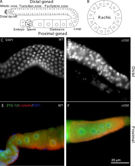
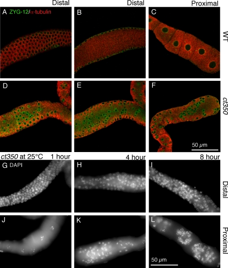
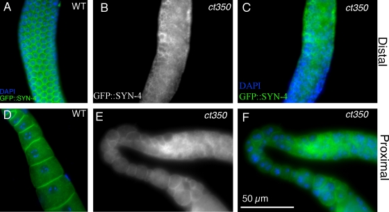
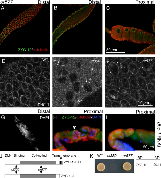
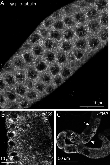
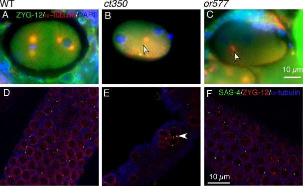
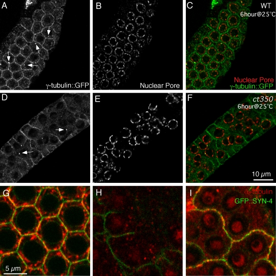
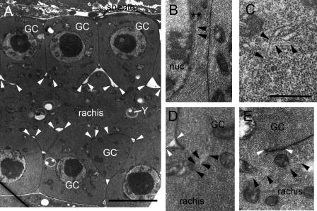
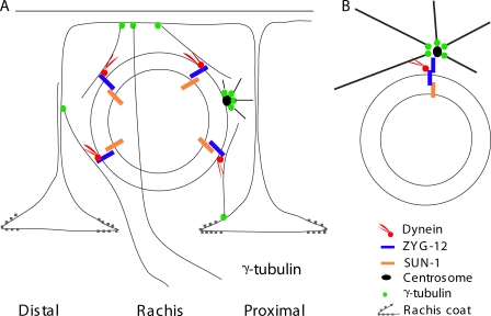
Changes in cellular microtubule organization often accompany developmental progression. In the Caenorhabditis elegans embryo, the centrosome, which is attached to the nucleus via ZYG-12, organizes the microtubule network. In this study, we investigate ZYG-12 function and microtubule organization before embryo formation in the gonad. Surprisingly, ZYG-12 is dispensable for centrosome attachment in the germline. However, ZYG-12-mediated recruitment of dynein to the nuclear envelope is required to maintain microtubule organization, membrane architecture, and nuclear positioning within the syncytial gonad. We examined gamma-tubulin localization and microtubule regrowth after depolymerization to identify sites of nucleation in germ cells. gamma-Tubulin localizes to the plasma membrane in addition to the centrosome, and regrowth initiates at both sites. Because we do not observe organized microtubules around zyg-12(ct350) mutant nuclei with attached centrosomes, we propose that gonad architecture, including membrane and nuclear positioning, is determined by microtubule nucleation at the plasma membrane combined with tension on the microtubules by dynein anchored at the nucleus by ZYG-12.


Figures

Figure 1.
Figure 1.
zyg-12(ct350ts) disrupts the regular arrangement of germline nuclei. (A and B) Schematic lateral (A) and transverse (B) view of germline nuclear position in the C. elegans gonad. (C and D) DAPI-stained germline nuclei are regularly arrayed in the distal gonad arm of adult wild-type animals (C) but disarrayed in zyg-12(ct350ts) gonads (D). (E and F) Immunofluorescent images with ZYG-12 in green, α-tubulin in red, and nuclei (DAPI) in blue. Oocytes contain a single nucleus in the proximal gonad of wild-type animals (E) but have multiple nuclei in zyg-12(ct350ts) gonads (F). All animals were shifted to 25°C during L1.
Figure 2.
Figure 2.
zyg-12(ct350) germline nuclei relocalize rapidly to the central rachis in response to a temperature shift. (A–F) Confocal images of gonads from egg-laying adults incubated at 25°C for 6 h. Gonads are labeled with ZYG-12 (green) to mark the nuclear envelope and α-tubulin (red). Wild-type (WT) gonads maintain the regular position of the nuclei at the periphery (A) and have no nuclei in the rachis (B). (C) Furthermore, the proximal gonads of wild-type animals have evenly spaced oocytes with one nucleus each. (D and E) In contrast, the nuclei at the periphery of zyg-12(ct350) gonads are disorganized (D), and many nuclei occupy the central rachis (E). (F) The proximal gonad of zyg-12(ct350) animals has abnormal clusters of small nuclei that resemble the distal nuclei. (G–L) DAPI-stained proximal (G–I) and distal (J–L) gonads of egg-laying adult zyg-12(ct350) animals incubated at 25°C for short intervals, as indicated. The nuclei are rapidly displaced in response to exposure to the restrictive temperature.
Figure 3.
Figure 3.
The membranes of zyg-12(ct350) gonads are rearranged at the restrictive temperature. (A–D) GFP::SYN-4 permits visualization of membranes in a wild-type distal (A) and proximal (D) gonad. This patterned array of germ cell membranes is disorganized in the distal (B and E) and proximal (C and F) arm of zyg-12(ct350) gonads.
Figure 4.
Figure 4.
The zyg-12(or577) mutant, which does not affect the ZYG-12–dynein interaction, has normal germline nuclear positioning. (A–C) Confocal fluorescent images of zyg-12(or577) incubated at 25°C for 6 h. Gonads are labeled with ZYG-12 (green) to mark the nuclear envelope and α-tubulin (red). (A and B) The nuclei at the periphery of the gonad maintain their regular positioning (A), and no nuclei are in the central rachis (B). (C) Furthermore, the proximal gonad has evenly spaced oocytes with one nucleus each, which is similar to wild-type animals (Fig. 2 C). The or577 allele has no detectable effect on ZYG-12–DLI-1 interactions in this assay. (D–F) Immunofluorescent images obtained with DHC-1 antibody stained gonads. (D) We confirmed that DHC-1 is recruited to the nuclear envelope of all nuclei in the wild-type (WT) germline. (E) In contrast, robust DHC-1 signal was never observed in zyg-12(ct350) mutant germlines (Malone et al., 2003), and <10% of the zyg-12(ct350) nuclei display a weak DLI-1 signal. (F) DHC-1 is retained at the nuclear envelope of all nuclei in zyg-12(or577) animals at 25°C. (G–I) RNAi of dynein heavy chain (dhc-1) disrupts gonad nuclear arrangement. (G) DAPI-stained distal gonad shows disorganized nuclei. (H and I) Immunofluorescent images of proximal gonads stained with ZYG-12 (green) to mark nuclei, α-tubulin (red), and DAPI (blue) show multiple nuclei (arrowhead) in single oocytes. (J) ct350 and or577 affect different domains of ZYG-12 (Malone et al., 2003). (K) We confirmed the ZYG-12 interaction with dynein light intermediate chain DLI-1 using a yeast two-hybrid assay as well as the lack of interaction caused by the ct350 allele (Malone et al., 2003). BD, DNA-binding domain vector; AD, activation domain vector.
Figure 5.
Figure 5.
Microtubule architecture accounts for nuclear positioning in both wild-type and ct350 mutants. (A) Distal gonad of wild-type (WT) animal. 3D projection of a z series of confocal immunofluorescent images stained with α-tubulin reveals a meshwork-like structure of microtubules surrounding the nuclei. Microtubules in the gonad of zyg-12(ct350) mutants are severely disorganized after 8 h at the restrictive temperature. (B) In a view of the distal gonad region, longer microtubule filaments crisscross through a cytoplasm devoid of nuclei. (C) In a view of the whole gonad arm, large microtubule networks encapsulate multiple nuclei. The arrowhead indicates multiple nuclei surrounded by the same microtubule network, and the arrow indicates nuclei not surrounded by microtubules.
Figure 6.
Figure 6.
There are different mechanisms controlling centrosome attachment in the embryo and the germline. (A–C) ZYG-12 is required for centrosomal attachment in the embryo. Immunofluorescent images of embryos incubated at the restrictive temperature for 6 h labeled with ZYG-12 (green) to mark the nuclear envelope and α-tubulin (red) to mark the centrosome. (A) The centrosomes are attached to the nuclei in a two-cell stage embryo. (B and C) Centrosomes (arrowheads) are detached from the nuclei in zyg-12(ct350) and zyg-12(or577) embryos. (D–F) ZYG-12 is dispensable for centrosome attachment in the gonad, and loss of nuclear anchoring is independent of centrosome attachment. Immunofluorescent images of gonads incubated at the restrictive temperature for 6 h labeled with antibodies to SAS-4 (green) to mark centrosomes, ZYG-12 (red) to mark nuclei, and α-tubulin (blue). Centrosomes remain closely apposed to the nuclear envelope of wild-type (WT) nuclei (D), zyg-12(ct350) nuclei that have been displaced (E), and zyg-12(or577) nuclei (F). Note that in wild-type and both mutants, the SAS-4 signal is visible at all nuclei but does not appear in images only because of the focal plane of the confocal image. Although nuclear position was disrupted, centrosomes still remain on the nuclear envelope of the zyg-12(ct350) mutants (E, arrowhead).
Figure 7.
Figure 7.
Plasma membrane as the nucleation site of microtubules in the distal gonad. (A–E) γ-Tubulin localizes to germ cell membrane in both wild-type (WT) and zyg-12(ct350) gonads. (A) In the distal gonad of wild-type animals, γ-tubulin localizes to germ cell membranes and centrosomes (white arrows). (B) The nuclear pore complex marks the nuclei. (D–F) In zyg-12(ct350), neither the membrane nor the centrosomal localization (D, white arrows) of γ-tubulin::GFP is altered. (F) However, nuclear position has been disrupted. (G–I) Microtubules regrow from the plasma membrane (marked by GFP::SYN-4) after the removal of depolymerizing drug from cultured gonads. (G) Filamentous microtubules occupy the cytoplasm and partially colocalize with the plasma membrane in a cultured gonad not subjected to nocodazole. (H) 1 h of nocodazole treatment effectively depolymerizes microtubules in the gonad. (I) 30 min after the removal of nocodazole, microtubules regrow at the plasma membrane.
Figure 8.
Figure 8.
TEM reveals microtubule organization in the germline. (A) Low power view of the distal arm of the gonad seen in lengthwise view. Rows of germ cells (GC) lie on either side of the rachis, connected by cytoplasmic bridges to the rachis. A somatic sheath cell partially covers the germline at this level. The cytoplasm of the germ cells contains a large central nucleus and scattered mitochondria. The rachis is also filled with mitochondria and contains yolk granules (Y) and some ER. Some organelles span the bridges between the germ cell and rachis. The outside surface of the rachis membrane has an electron-dense rachis coat between closely spaced arrowheads. (B–E) Higher power views of germ cells oriented so that the germ cell lies to the top of each panel with the rachis beneath it, running left to right as in A. Note that free ribosomes fill most of the cytoplasm in all distal germline cells and in the rachis. (B) A microtubule appears to attach to an electron-dense projection (asterisks) on the cytoplasmic face of a germ cell nucleus (nuc). These projections appear to extend from the nuclear pore. (C) Two microtubules pass one another at almost orthogonal angles near the top of a germ cell. (D) A bundle of microtubules passes across the cytoplasmic bridge between the rachis and a germ cell, running close to several mitochondria. (E) Microtubules can be seen running lengthwise along the long axis of the rachis close to several mitochondria. Black arrowheads indicate microtubules, and white arrowheads mark rachis openings for each germ cell. Bars: (A) 5 µm; (C) 0.5 µm.
Figure 9.
Figure 9.
A model for MTOC-independent nuclear anchoring in the germline. (A) A model for germline nuclear anchoring in the gonad. ZYG-12 recruits dynein to the outer nuclear envelope. Dynein promotes nuclear anchoring via direct interactions with the microtubule filaments nucleated from membrane-localized γ-tubulin. The centrosome is not involved in nuclear positioning of germline nuclei. (B) The centrosome is the key to nuclear positioning in the embryo. ZYG-12 homodimerization is essential to maintain the nucleus–centrosome interaction in the embryo, permitting control of nuclear positioning.

Similar articles

Cited by

References

    1. Baas P.W., Deitch J.S., Black M.M., Banker G.A. 1988. Polarity orientation of microtubules in hippocampal neurons: uniformity in the axon and nonuniformity in the dendrite.Proc. Natl. Acad. Sci. USA. 85:8335–8339 - PMC - PubMed
    1. Bre M.H., Pepperkok R., Hill A.M., Levilliers N., Ansorge W., Stelzer E.H., Karsenti E. 1990. Regulation of microtubule dynamics and nucleation during polarization in MDCK II cells.J. Cell Biol. 111:3013–3021 - PMC - PubMed
    1. Brenner S. 1974. The genetics of Caenorhabditis elegans.Genetics. 77:71–94 - PMC - PubMed
    1. Bugnard E., Zaal K.J., Ralston E. 2005. Reorganization of microtubule nucleation during muscle differentiation.Cell Motil. Cytoskeleton. 60:1–13 - PubMed
    1. Dammermann A., Muller-Reichert T., Pelletier L., Habermann B., Desai A., Oegema K. 2004. Centriole assembly requires both centriolar and pericentriolar material proteins.Dev. Cell. 7:815–829 - PubMed

Publication types

MeSH terms