. 2009 Aug 3;206(8):1755-67.
doi: 10.1084/jem.20082118. Epub 2009 Jul 27.

Negative feedback control of the autoimmune response through antigen-induced differentiation of IL-10-secreting Th1 cells

Affiliations

Negative feedback control of the autoimmune response through antigen-induced differentiation of IL-10-secreting Th1 cells

Leona Gabrysová et al. J Exp Med. .

Abstract

Regulation of the immune response to self- and foreign antigens is vitally important for limiting immune pathology associated with both infections and hypersensitivity conditions. Control of autoimmune conditions can be reinforced by tolerance induction with peptide epitopes, but the mechanism is not currently understood. Repetitive intranasal administration of soluble peptide induces peripheral tolerance in myelin basic protein (MBP)-specific TCR transgenic mice. This is characterized by the presence of anergic, interleukin (IL)-10-secreting CD4(+) T cells with regulatory function (IL-10 T reg cells). The differentiation pathway of peptide-induced IL-10 T reg cells was investigated. CD4(+) T cells became anergic after their second encounter with a high-affinity MBP peptide analogue. Loss of proliferative capacity correlated with a switch from the Th1-associated cytokines IL-2 and interferon (IFN)-gamma to the regulatory cytokine IL-10. Nevertheless, IL-10 T reg cells retained the capacity to produce IFN-gamma and concomitantly expressed T-bet, demonstrating their Th1 origin. IL-10 T reg cells suppressed dendritic cell maturation, prevented Th1 cell differentiation, and thereby created a negative feedback loop for Th1-driven immune pathology. These findings demonstrate that Th1 responses can be self-limiting in the context of peripheral tolerance to a self-antigen.


Figures

Figure 1.
Figure 1.
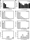
Changes in serum cytokine profile over the course of tolerance induction. Tg4 Rag1−/− mice were bled before treatment and after each successive i.n. MBP Ac1-9[4Y] administration. Serum concentrations of IL-2, IL-10, IFN-γ, IL-17, IL-4, and IL-5 were measured by cytometric bead assay. Results show the mean serum cytokine concentration of six to eight mice per treatment group on pooled data from two separate experiments.
Figure 2.
Figure 2.
Anergy-associated down-regulation of Th1 cytokines and increase in IL-10 production by CD4+ T cells during tolerization. Splenic CD4+ T cells were positively selected from untreated or i.n. MBP Ac1-9[4Y]–treated Tg4 Rag1−/− mice at 2 h after each peptide administration. 5 × 10⁴ CD4+ T cells per well were cultured with 100 µg/ml of MBP Ac1-9[4K] in the presence of 105 autologous irradiated CD4 depleted splenocytes as APCs and, where indicated, 20 U/ml of rhIL-2. (A and B) Proliferative responses were measured at 72 h by 3[H]thymidine incorporation in the absence or presence of rhIL-2. Results are depicted as the mean thymidine incorporation ± SEM. (C and D) Cytokine responses of CD4+ T cells from the cultures described in A and B were measured by ELISA at 24 h (IL-2) and 72 h (IFN-γ and IL-10) after in vitro restimulation in the absence or presence of rhIL-2. Results are depicted as the mean cytokine production in ng/ml of supernatant ± SEM. Data in each panel are representative of three individual experiments.
Figure 3.
Figure 3.
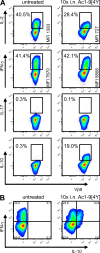
Th1 differentiation pathway of IL-10 T reg cells from tolerized mice. Tg4 Rag1−/− mice were treated with a minimum of 10 i.n. doses of MBP Ac1-9[4Y]. Splenic CD4+ T cells were positively selected from either untreated or peptide-treated mice 3 d after the last treatment and cultured with 10 µg/ml of MBP Ac1-9[4K] in the presence of irradiated B10.PL splenocytes as APCs. RhIL-2 (20 U/ml) was added on d 1 and 3 to CD4+ T cell cultures from peptide-treated and untreated mice, respectively. (A and B) Intracellular cytokine staining for IL-2, IFN-γ, IL-17, and IL-10 was performed on d 7 of culture after additional restimulation with PMA and ionomycin. Depicted are density plots of Vβ8+ T cells with cytokine gates based on isotype controls. Data are representative of at least three individual experiments.
Figure 4.
Figure 4.
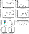
Expression of genes associated with differentiation of IL-10 T reg cells. (A–D) Tg4 Rag1−/− mice were treated with a minimum of 10 i.n. doses of MBP Ac1-9[4Y]. Total RNA was extracted from splenic CD4+ T cells positively selected from either untreated or peptide-treated mice at 2 h after each peptide treatment. The RNA was reverse transcribed and expression levels of the transcription factors t-bet, gata-3, egr-2, and the co-stimulatory molecule icos were determined by quantitative real-time PCR after normalization to β2-microglobulin expression. Results show the relative expression of the gene of interest from three accumulated experiments at each time point. (E) Enriched IL-10–secreting CD4+ T cells from Tg4 mice treated with a minimum of 10 i.n. doses of MBP Ac1-9[4Y] and in vitro–differentiated Th1 and Th2 cells were stained for surface Vβ8 and intracellular T-bet (black line) or isotype control (gray line). The depicted density plot of IL-10–enriched CD4+ T cells is gated on Vβ8+ T cells. The histogram of the IL-10+ cell fraction is gated on Vβ8+IL-10+ cells and Th1/Th2 cell histograms on Vβ8+ T cells. Data are representative of at least three individual experiments. (F) FACS-sorted IL-10+, IL-10+IFN-γ+, and IL-10+IFN-γ Tg4 CD4+ T cells were analyzed for T-bet expression by Western blot, with Th1 and naive (CD4+CD62L+) cells used as positive and negative controls for T-bet detection, respectively. Data are representative of two individual experiments.
Figure 5.
Figure 5.
Role of IL-10 in down-regulation of Th1 cytokines in tolerance. Tg4 and Tg4 IL-10−/− mice were treated with a minimum of 10 i.n. doses of MBP Ac1-9[4Y]. Splenic CD4+ T cells were positively selected from either untreated Tg4 or peptide-treated Tg4 and Tg4 IL-10−/− mice 3 d after the last peptide treatment. 5 × 10⁴ CD4+ T cells per well were cultured with 105 irradiated B10.PL splenocytes as APCs in the presence of a 10-fold titration of MBP Ac1-9[4K], ranging from 0.1 to 100 µg/ml. (A) Proliferative responses were measured at 72 h by 3[H]thymidine incorporation. Results are depicted as the mean thymidine incorporation ± SEM. (B–D) Cytokine responses of CD4+ T cells from the above cultures were measured by ELISA at 24 h (IL-2) and 72 h (IFN-γ and IL-10) after in vitro restimulation. Results are expressed as the mean cytokine production in nanogram/milliliter of supernatant ± SEM. Data are representative of at least three individual experiments.
Figure 6.
Figure 6.
Redundancy of non-T cell–derived IL-10 in IL-10 T reg cell differentiation. Negatively selected Tg4 CD4+ T cells at 2 × 10⁷ per mouse were adoptively transferred into either B10.PL or B10.PL IL-10−/− recipients. 7 d later, mice were treated with a minimum of 10 i.n. doses of MBP Ac1-9[4Y] at 3–4 d intervals or left untreated as controls. Spleens from peptide-treated recipient mice were collected 3 d after the last peptide treatment. Tg4 CD4+ T cells were selected by culturing with 10 µg/ml of Ac1-9[4K] and rhIL-2 for 7 d and stained for surface Vβ8 and intracellular IFN-γ or IL-10 after additional restimulation with PMA and ionomycin. The depicted contour plots are gated on Vβ8+ T cells with quadrants based on isotype controls. Data are representative of at least three individual experiments.
Figure 7.
Figure 7.
IL-10R signaling requirement for IL-10 T reg cell–mediated suppression of DC function. Tg4 and Tg4 IL-10−/− mice were treated with a minimum of 10 i.n. doses of MBP Ac1-9[4Y]. Splenic DCs were isolated from untreated B10.PL mouse spleens and 5 × 10⁴ DCs were cultured either alone or with equal numbers of CD4+ T cells positively selected from untreated or peptide-treated Tg4 or Tg4 IL-10−/− mice. 100 µg/ml of Ac1-9[4K] and/or 10 µg/ml of anti–IL-10R or isotype control antibody were added where indicated. (A) After 24 h, DCs were stained for surface CD11c, CD80, CD86, and CD40. Results are depicted histograms for each surface protein after gating on CD11c+ cells, with x and y axes showing the fluorescence intensity and percentage of max, respectively. (B) Supernatant cultures described in A were analyzed for IL-12 by ELISA at 24 h after in vitro restimulation. Results are depicted as the mean IL-12 production in nanogram/milliliter of supernatant ± SEM. Data are representative of at least two individual experiments.
Figure 8.
Figure 8.
Role of IL-10 in negative feedback regulation of Th1 responses. Tg4 mice were treated with a minimum of 10 i.n. doses of MBP Ac1-9[4Y]. Splenic DCs were isolated from both untreated and peptide-treated Tg4 mice that were administered 100 µg of MBP Ac1-9[4Y] 3 h previously. CD4+ T cells positively selected from untreated Tg4 mouse spleens were cultured at 5 × 10⁴ per well in the presence of a twofold titration of irradiated DCs at a ratio ranging from 80:1 to 10:1 and 10 µg/ml of Ac1-9[4K]. (A) Proliferative responses were measured at 72 h by 3[H]thymidine incorporation. Results are depicted as the mean thymidine incorporation ± SEM. (B) Cytokine responses of CD4+ T cell from the 10:1 cultures were measured by ELISA at 24 h (IL-2) and 72 h (IFN-γ and IL-12) after in vitro restimulation. Results are depicted as the mean cytokine production in ng/ml of supernatant ± SEM. Data are representative of two individual experiments.

References

    1. Adler H.S., Kubsch S., Graulich E., Ludwig S., Knop J., Steinbrink K. 2007. Activation of MAP kinase p38 is critical for the cell-cycle-controlled suppressor function of regulatory T cells.Blood. 109:4351–4359 - PubMed
    1. Anderson C.F., Oukka M., Kuchroo V.J., Sacks D. 2007. CD4(+)CD25(-)Foxp3(-) Th1 cells are the source of IL-10-mediated immune suppression in chronic cutaneous leishmaniasis.J. Exp. Med. 204:285–297 - PMC - PubMed
    1. Anderson P.O., Manzo B.A., Sundstedt A., Minaee S., Symonds A., Khalid S., Rodriguez-Cabezas M.E., Nicolson K., Li S., Wraith D.C., et al. 2006. Persistent antigenic stimulation alters the transcription program in T cells, resulting in antigen-specific tolerance.Eur. J. Immunol. 36:1374–1385 - PMC - PubMed
    1. Anderson P.O., Sundstedt A., Yazici Z., Minaee S., O'Neill E.J., Woolf R., Nicolson K., Whitley N., Li L., Li S., et al. 2005. IL-2 overcomes the unresponsiveness but fails to reverse the regulatory function of antigen-induced T regulatory cells.J. Immunol. 174:310–319 - PubMed
    1. Andris F., Denanglaire S., de Mattia F., Urbain J., Leo O. 2004. Naive T cells are resistant to anergy induction by anti-CD3 antibodies.J. Immunol. 173:3201–3208 - PubMed

Publication types

MeSH terms