. 2009 Aug 3;206(8):1787-802.
doi: 10.1084/jem.20090641. Epub 2009 Jul 27.

Myocyte necrosis underlies progressive myocardial dystrophy in mouse dsg2-related arrhythmogenic right ventricular cardiomyopathy

Affiliations

Myocyte necrosis underlies progressive myocardial dystrophy in mouse dsg2-related arrhythmogenic right ventricular cardiomyopathy

Kalliopi Pilichou et al. J Exp Med. .

Abstract

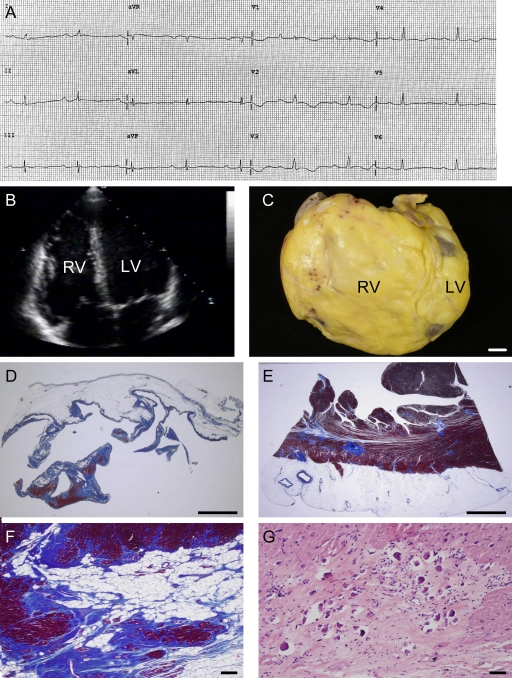
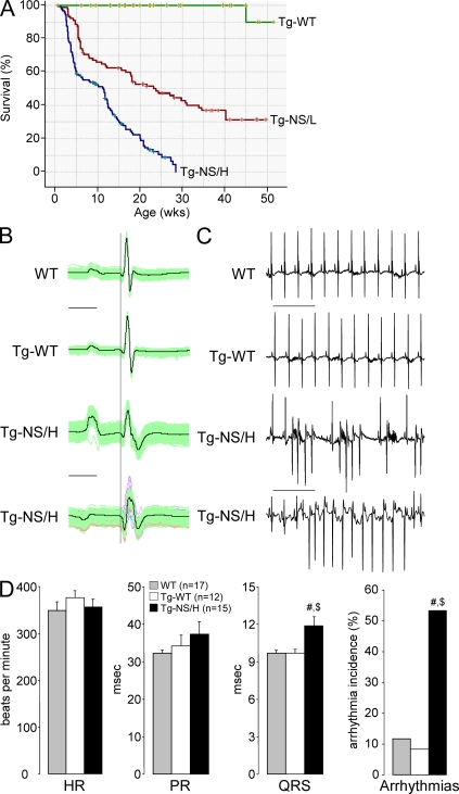
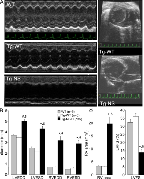
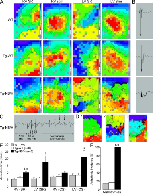
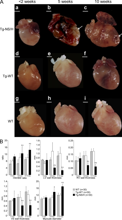
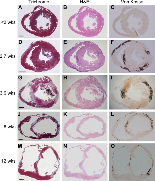
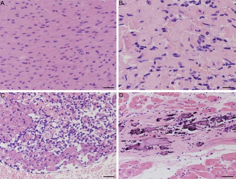
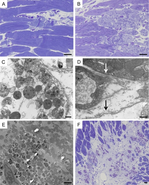
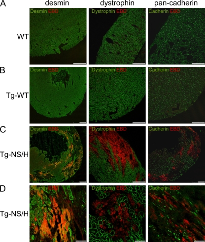
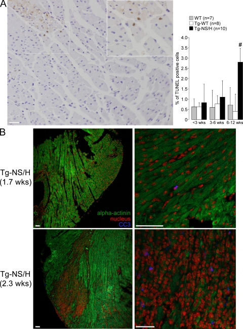
Mutations in the cardiac desmosomal protein desmoglein-2 (DSG2) are associated with arrhythmogenic right ventricular cardiomyopathy (ARVC). We studied the explanted heart of a proband carrying the DSG2-N266S mutation as well as transgenic mice (Tg-NS) with cardiac overexpression of the mouse equivalent of this mutation, N271S-dsg2, with the aim of investigating the pathophysiological mechanisms involved. Transgenic mice recapitulated the clinical features of ARVC, including sudden death at young age, spontaneous ventricular arrhythmias, cardiac dysfunction, and biventricular dilatation and aneurysms. Investigation of transgenic lines with different levels of transgene expression attested to a dose-dependent dominant-negative effect of the mutation. We demonstrate for the first time that myocyte necrosis is the key initiator of myocardial injury, triggering progressive myocardial damage, including an inflammatory response and massive calcification within the myocardium, followed by injury repair with fibrous tissue replacement, and myocardial atrophy. These observations were supported by findings in the explanted heart from the patient. Insight into mechanisms initiating myocardial damage in ARVC is a prerequisite to the future development of new therapies aimed at delaying onset or progression of the disease.


Figures

Figure 1.
Figure 1.
Characteristics of the female patient carrier of the DSG2-N266S mutation who underwent heart transplantation at the age of 65. (A) 12-lead ECG 2 mo before heart transplantation displaying sinus rhythm with PQ and QT prolongation, intraventricular conduction delay, and negative T wave V1–V4. (B) Two-dimensional echocardiography showing biventricular dilatation. (C) Gross view of the explanted heart specimen showing massive RV dilatation with RV outflow tract aneurysm. Bar, 1 cm. (D and E) RV and LV free wall showing fibro-fatty replacement of the myocardium with almost total disappearance of the myocardium in the RV. Bar, 5 mm. (F) Close-up of E. Bar, 30 µm. (G) At higher magnification, spotty calcification is observed in areas of replacement-type fibrosis. Bar, 50 µm.
Figure 2.
Figure 2.
Characteristics of transgenic mice with cardiac-restricted expression of N271S-dsg2. (A) Kaplan-Meier survival plot of Tg-NS/H (blue line; n = 225), Tg-NS/L (red line; n = 156), and Tg-WT (green line; n = 129) mice. Note the high death rate in the mutant lines, with 30% of death occurring by 3.6 wk in Tg-NS/H mice. (B) Examples of surface ECG measurements from WT, Tg-WT, and Tg-NS/H mice showing prolongation and fractionation of the QRS complex, indicating impairment of ventricular conduction. Bar, 25 ms. (C) Typical ECG examples of spontaneous ventricular arrhythmias observed in Tg-NS/H mice. Bar, 500 ms. (D) Mean values of ECG parameters including heart rate (HR), PR and QRS intervals, and incidence of spontaneous arrhythmias. Data represent means ± SEM measured from >200 consecutive beats from single independent ECG measurements in 17 WT, 12 Tg-WT, and 15 Tg-NS/H mice (#, P < 0.05 vs. WT; $, P < 0.05 vs. Tg-WT; Table S1).
Figure 3.
Figure 3.
Assessment of in vivo cardiac morphology and function. (A) M-mode and two-dimensional echocardiography in WT, Tg-WT, and Tg-NS/H mice displaying LV and RV chamber dilatation in mutant mice. (B) Mean values of echocardiographic parameters from WT, Tg-WT, and Tg-NS/H mice, including LV and RV end-diastolic diameter (LVEDD and RVEDD, respectively), LV and RV end-systolic diameter (LVESD and RVESD, respectively), RV area (RV area in diastole [short-axis view]), and LV fractional shortening (LVFS). Data represent means ± SEM from single independent echocardiographic measurements on five mice per experimental group (#, P < 0.05 vs. WT; *, P < 0.001 vs. WT; $, P < 0.01 vs. Tg-WT; &, P < 0.001 vs. Tg-WT; Table S3).
Figure 4.
Figure 4.
Epicardial mapping experiments in Langendorff-perfused hearts. (A) Examples of RV and LV activation maps for WT, Tg-WT, and Tg-NS/H hearts during sinus rhythm (SR) and stimulation from the center of the electrode (stim). Crowding of isochrones in Tg-NS/H hearts indicates areas of conduction slowing. (B) Examples of extracellular electrograms showing fractionation of the extracellular signal (a sign of excessive conduction slowing) in Tg-NS/H but not in WT or Tg-WT mice. (C) Induction of VT in a Tg-NS/H heart after administration of two short-coupled extrastimuli (S1 and S2). (D) Ventricular activation map during beats 1–3 of the VT depicted in C. (E) Mean RV and LV activation times during SR and central stimulation (CS). (F) Mean incidence of inducible arrhythmias. Data represent means ± SEM from single independent epicardial mapping experiments from five WT, six Tg-WT, and five Tg-NS/H mice (#, P < 0.05 vs. WT; ‡, P < 0.01 vs. WT; $, P < 0.05 vs. Tg-WT; Table S4).
Figure 5.
Figure 5.
Gross features of explanted mouse hearts. (A) Tg-NS/H, Tg-WT, and WT hearts at age <2 (a, d, and g), 5 (b, e, and h), 10 (c, f, and i) wk. Note the structurally normal hearts at young age, with later appearance of calcification and dilatation with aneurysms (arrows) in Tg-NS/H hearts. Bars: (a, b, and d–i) 1 mm; (c) 2 mm. (B) Mean values of HW/BW ratios; LV, RV, and ventricular septum (VS) wall thicknesses; and cardiomyocyte diameters in hearts of different age groups (<3, 3–6, and 6–12 wk; 10 mice per age group for each line; #, P < 0.01 vs. WT; ‡, P < 0.01 vs. Tg-WT; Table S5).
Figure 6.
Figure 6.
Histological features of explanted hearts from Tg-NS/H mice. (A–O) Heidenhain trichrome, H&E, and von Kossa staining of transverse full sections of the hearts at different ages: <2 (A–C), 2.7 (D–F), 3.6 (G–I), 8 (J–L), and 12 (M–O) wk. Note the appearance of myocardial damage in the outer subepicardial layers with calcification in both RV and LV free walls after 2 wk of age, with progressive extension to become transmural by 4 wk of age, and deposition of mature fibrosis with wall thinning and aneurysm formation by 8 wk of age. Bars: (A–C) 0.5 mm; (D–F) 0.8 mm; (G–I) 0.5 mm; (J–L) 0.4 mm; and (M–O) 0.8 mm.
Figure 7.
Figure 7.
Histological features of Tg-NS/H hearts at high magnification. (A) Normal myocardium (1.7 wk of age). Bar, 50 µm. (B) Cardiomyocyte necrosis with early neutrophil infiltrates (2.1 wk of age). Bar, 20 µm. (C) Diffuse inflammatory cells including macrophages (3.6 wk of age). Bar, 50 µm. (D) Replacement-type fibrosis with spots of calcification (8 wk of age). Bar, 30 µm.
Figure 8.
Figure 8.
Ultrastructural features of cardiomyocyte necrosis in Tg-NS/H mice. (A) Focal contraction band necrosis on a semithin section. Bar, 10 µm. (B) Myocytolysis on a semithin section. Bar, 10 µm. (C) Disgregation of cytoplasm with loss of myofilaments and woolly bodies inside swollen mitochondria (m), features in keeping with necrotic cell death. Bar, 500 nm. (D) Loss of sarcolemma integrity in a cardiomyocyte (black arrow) with cytoplasmic disgregation, close to abnormal cardiomyocyte with intact sarcolemma (white arrow). Bar, 500 nm. (E) Mineralization of mitochondria. Bar, 2 µm. (F) Spotty calcification within fibrous tissue on a semithin section. Bar, 10 µm.
Figure 9.
Figure 9.
Immunohistochemical analysis of cardiac cryosections from 2–3-wk-old mice injected with EBD. (A and B) WT and Tg-WT hearts show normal distribution of desmin, dystrophin, and pan-cadherin in the ventricles, and no EBD-positive cells. Bars, 250 µm. (C and D) In contrast, hearts from Tg-NS mice display large areas of ventricular myocardium lacking desmin, dystrophin, and pan-cadherin. These abnormal areas contained myocytes positive for EBD, indicating increased cell membrane permeability and loss of myocyte integrity and viability. Bars: (C) 250 µm; (D) 50 µm.
Figure 10.
Figure 10.
Cardiomyocyte apoptosis in Tg-NS/H mice. (A, left) TUNEL analysis of cardiac apoptosis. Note the presence of positive-stained myocyte nuclei (brown) close to an area of myocyte necrosis. (inset) Close-up of boxed area. Bar, 50 µm. (right) Mean values are depicted for the percentage of TUNEL-positive cells in three age groups. Data in this figure are representative of 7 WT, 8 Tg-WT, and 10 Tg-NS/H mice. #, P < 0.05 vs. WT and Tg-WT. (B) Immunohistochemical analysis of cleaved caspase-3 (CC3) for assessment of apoptosis in cardiac cryosections from Tg-NS mice. In Tg-NS mice <2 wk old (1.7 wk old in this example), cardiac structure was still unaltered, as indicated by the normal α-actinin staining pattern, and CC3-positive cells were hardly observed. In contrast, in hearts of Tg-NS mice of >2 wk old, clear CC3 staining was observed, but only in necrotic areas of the ventricular myocardium. Bars, 50 µm.

References

    1. Asimaki A., Syrris P., Wichter T., Matthias P., Saffitz J.E., McKenna W.J. 2007. A novel dominant mutation in plakoglobin causes arrhythmogenic right ventricular cardiomyopathy.Am. J. Hum. Genet. 81:964–973 - PMC - PubMed
    1. Bansal D., Miyake K., Vogel S.S., Groh S., Chen C.C., Williamson R., McNeil P.L., Campbell K.P. 2003. Defective membrane repair in dysferlin-deficient muscular dystrophy.Nature. 423:168–172 - PubMed
    1. Basso C., Thiene G., Corrado D., Angelini A., Nava A., Valente M. 1996. Arrhythmogenic right ventricular cardiomyopathy. Dysplasia, dystrophy, or myocarditis? Circulation. 94:983–991 - PubMed
    1. Basso C., Tsatsopoulou A., Thiene G., Anastasakis A., Valente M., Protonotarios N. 2001. “Petrified” right ventricle in long-standing naxos arrhythmogenic right ventricular cardiomyopathy.Circulation. 104:E132–E133 - PubMed
    1. Basso C., Czarnowska E., Della Barbera M., Bauce B., Beffagna G., Wlodarska E.K., Pilichou K., Ramondo A., Lorenzon A., Wozniek O., et al. 2006. Ultrastructural evidence of intercalated disc remodelling in arrhythmogenic right ventricular cardiomyopathy: an electron microscopy investigation on endomyocardial biopsies.Eur. Heart J. 27:1847–1854 - PubMed

Publication types

MeSH terms