

Comparative Study
doi: 10.1161/CIRCULATIONAHA.108.796698.
Human cardiac development in the first trimester: a high-resolution magnetic resonance imaging and episcopic fluorescence image capture atlas
Affiliations
- PMID: 19635979
- PMCID: PMC3411176
- DOI: 10.1161/CIRCULATIONAHA.108.796698
Item in Clipboard
Comparative Study
Human cardiac development in the first trimester: a high-resolution magnetic resonance imaging and episcopic fluorescence image capture atlas
Circulation.
.
No abstract available
Figures

2D EFIC image stacks were reconstructed in 3D to show the looped heart tube in a EGA 7 5/7 weeks (CS17) embryo. The double headed arrow indicates the interventricular foramen. The orifice of the developing atrioventricular junction is seen as a horizontal line above the label AV. The truncus arteriosus (arrowhead) is also seen. Endocardial cushion tissue surrounding the atrioventricular junction is adjacent to the truncus arteriosus. RA: right atrium, LA: left atrium, RV: presumptive right ventricle, LV: presumptive left ventricle. Scale bar = 0.6 mm.
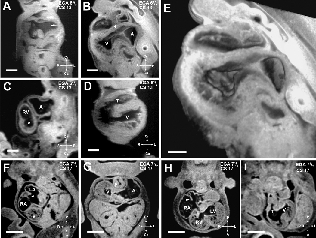
(A–E). EFIC and MRI images of EGA 6 4/7 weeks (CS13) embryos shown in various imaging planes. Imaging in the frontal plane (A) shows the common cardinal veins or the open venous confluence (arrow), while sagittal view (B) shows primitive endocardial cushions at the atrioventricular junction (arrowhead). A 3D model of the same embryo (E) shows the extent of the interventricular foramen as well as the contour of the endocardial cushions. MRI image of another embryo in the sagittal plane (C) shows the presumptive right ventricle (RV), atrial chamber (A), and a nondistinct interventricular foramen (*), while the ventricular chamber (V) and a single, undivided truncus arteriosus (T) can be seen in an frontal section of a third embryo (D). Scale bars: (A–D)=0.4 mm, (E)=0.25 mm (F–I). MRI images of EGA 7 5/7 weeks (CS17) embryo. Image from an oblique transverse plane (F) shows the right and left atrial (RA, LA) chambers as septation is progressing (arrowhead). The developing ventricle (V) is seen. Viewed in the transverse plane in (G), well formed dense endocardial cushion tissue is seen at the atrioventricular junction (arrowhead). Another section in the transverse plane in (H) shows the right and left ventricular cavities with a more distinct interventricular foramen (*). Septum primum can be seen as atrial septation progresses (arrowhead). The single undivided truncus arteriosus (T) and interventricular foramen communicating with the presumptive left ventricle (V) can be seen in an oblique transverse plane (I). Scale bar: (E–H) = 1.250 mm. LV: presumptive left ventricular chamber, T: truncus arteriosus.
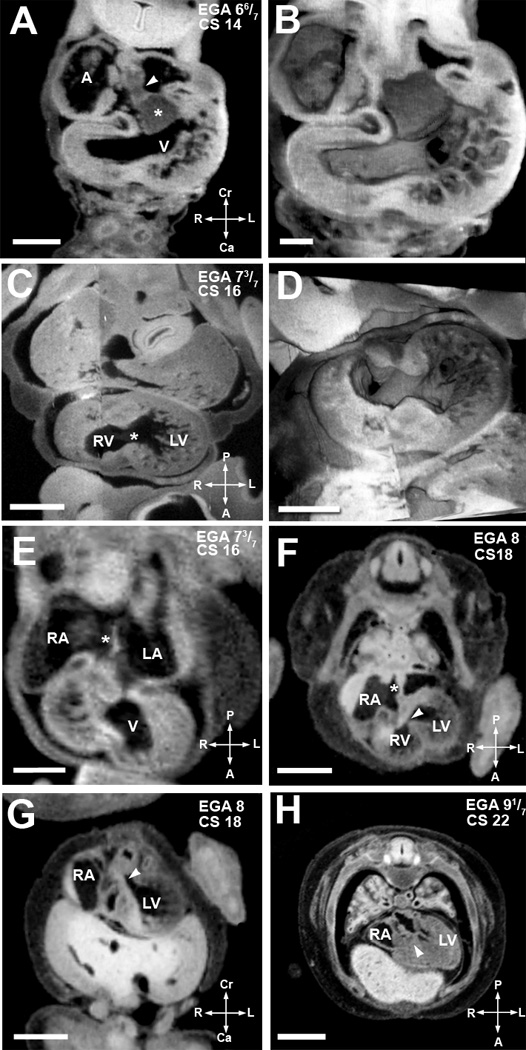
(A,B). EFIC image of EGA 6 6/7 weeks (CS14) embryo in the transverse plane (A) shows the atrial spine (arrowhead) attached to the inferior cushion (asterisks). 3D reconstruction (B) highlights the endocardial cushions and trabeculation in the ventricular chamber. Scale bar = 0.515 mm in (A), 0.272mm in (B). (C,D). An EFIC image of EGA 7 3/7 weeks (CS16) embryo in the oblique plane (C) showing right and left ventricular chambers connected by an interventricular foramen (*). 3D reconstruction of the same embryo (D) delineates the contour of the interventricular foramen and the orifices of the atrioventricular canal and the truncus arteriosus. Scale bar = 0.5mm for (C), and 0.900mm for (D). (E) MRI image of an embryo at EGA 7 3/7 weeks (CS16) also in the transverse plane. It shows the formation of septum primum (*) between the right and left atria (RA, LA). Scale bar = 0.5 mm. (F,G) MRI image of an EGA 8 weeks (CS18) embryo in an oblique transverse plane (F) shows a complete atrial septum (*). The most caudal portion of the septum primum, the mesenchymal cap, has fused to the superior cushion. The growth of the muscular ventricular septum into the ventricular cavity is also shown. The crest of the muscular interventricular septum is present with an incomplete inlet ventricular septum (arrowhead) immediately above it. Panel G, another MRI image of the same embryo in an oblique coronal plane, shows the formed outlet ventricular septum (arrowhead). Together these two images show that outlet ventricular septation is completed before inlet ventricular septation. Scale bars in (F,G) = 1.5 mm. (H). MRI image of embryo at EGA 9 1/7 weeks (CS22) in an oblique coronal plane shows a completed inlet ventricular septum (arrowhead). Scale bar = 2mm. A: primitive atrium/venous confluence, RA: right atrium, LA: left atrium, V: ventricular chamber, LV: left ventricular chamber, RV: right ventricular chamber.
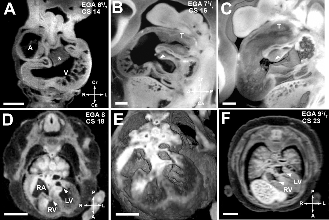
(A). EFIC image of an embryo at EGA 6 6/7 weeks (CS 14) in the transverse plane shows a large endocardial cushion (*) in the center of the cardiac loop. Scale bar = 0.515 mm (B,C). EFIC image of an embryo at EGA 7 3/7 weeks (CS16) in a sagittal plane (B) shows a tight, well formed atrioventricular junction (arrowhead) and the truncus arteriosus (T). 3D volume of the same embryo (C) shows exquisite detail of the contour and shape of the endocardial cushions and the truncus arteriosus (T). Scale bar in (B,C)=0.389mm. (D,E). MRI image in an oblique transverse plane (D) of an embryo at EGA 8 weeks (CS 18) shows separate atrioventricular valves. The valve leaflets appear thick at this stage. Note right and left atrioventricular valves denoted by arrowheads. 3D volume of the same embryo (E) shows indentation associated with the opening in the inlet ventricular septum. Scale bar for (D)=1.5 mm, (E)=1.1mm. (F) MRI image in an oblique transverse plane of a more mature EGA 9 3/7 weeks (CS 23) embryo. It shows separate atrioventricular valves with thinner valve leaflets (arrowheads). The inlet septum is closed. Scale bar = 2 mm. A: primitive atrium, RA: right atrium, V: ventricular chamber, RV: right ventricular chamber, LV: left ventricular chamber
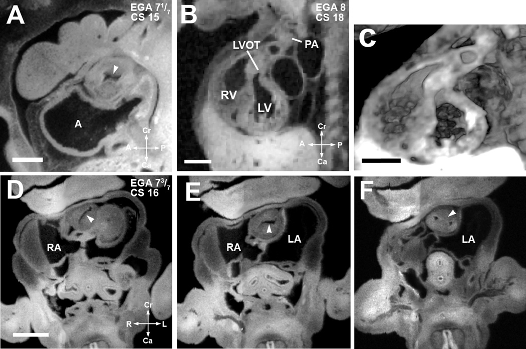
(A). EFIC image of an embryo at EGA 7 1/7 weeks (CS 15) in the sagittal plane shows a single orifice of the truncus arteriosus with inward swelling of the aorticopulmonary septum (arrowhead) which precedes septation of the truncus arteriosus. Scale bar = 0.622 mm. (B,C) EFIC image of an EGA 8 (CS 18) embryo (B) show a distinct pulmonary artery (PA) emerging from the right ventricle (RV) and a left ventricular outflow tract (LVOT) or aorta emerging from the left ventricle (LV). 3D volume of the same embryo (C) shows crossing of the great arteries. Scale bar in (B,C)=1.35 mm. (D–F). EFIC images of a embryo at EGA 7 3/7 weeks (CS 16) in oblique transverse planes showing the truncus arteriosus. Note changing orientation of the lumen (D,E) indicative of spiraling of the cushions (see arrowheads). In (F), the aorticopulmonary septum (arrowhead) has divided the distal portion of the truncus arteriosus into two separate arterial channels to the right and left of the aorticopulmonary septum. Scale bar = 0.9 mm. A: primitive atrium, RA: right atrium, LA: left atrium, RV: right venetricle, LV: left ventricle.

Outlined in the chart is the timing for major cardiac morphogenetic events and the presence of various cardiac structures in the human embryo. The timeline indicated for Atrioventricular Junction/Valve Formation (green bar) refer to when a distinct atrioventricular junction is observed before atrioventricular valve leaflets are evident. The timelime indicated for Semilunar Valve Formation (orange bar) refer to when distinct truncal cushion tissue is observed and before semilunar valve leaflets are evident. The demarcation of Mitral Valve, Tricuspid Valve, Aortic Valve and Pulmonary Valve delineate the developmental stages when distinct valve leaflets are observed and the stages when the valve leaflets continue to undergo maturation and thinning. The timeline indicated for Interventricular Foramen refer to when any communication is present between the right and left ventricular chambers.
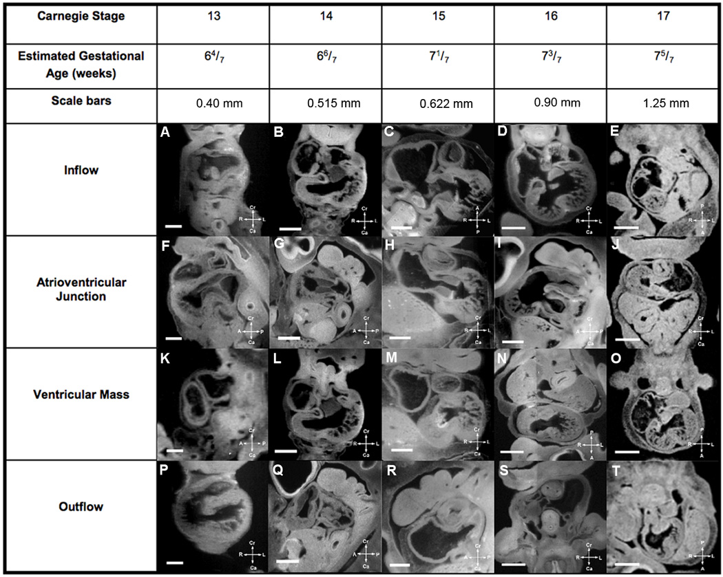
Major cardiac developmental structures present in EGA 6 4/7 weeks to 7 5/7 weeks (CS13–17) embryos are summarized in Figure 7, while Figure 8 shows the major developmental structures present in EGA 8 weeks to 9 3/7 weeks (CS18–23) embryos. One can look at a developmental structure at a specific estimated gestational age to determine what normal development is for that structure at that age in human cardiac development. The compass orients the observer to the plane of section.
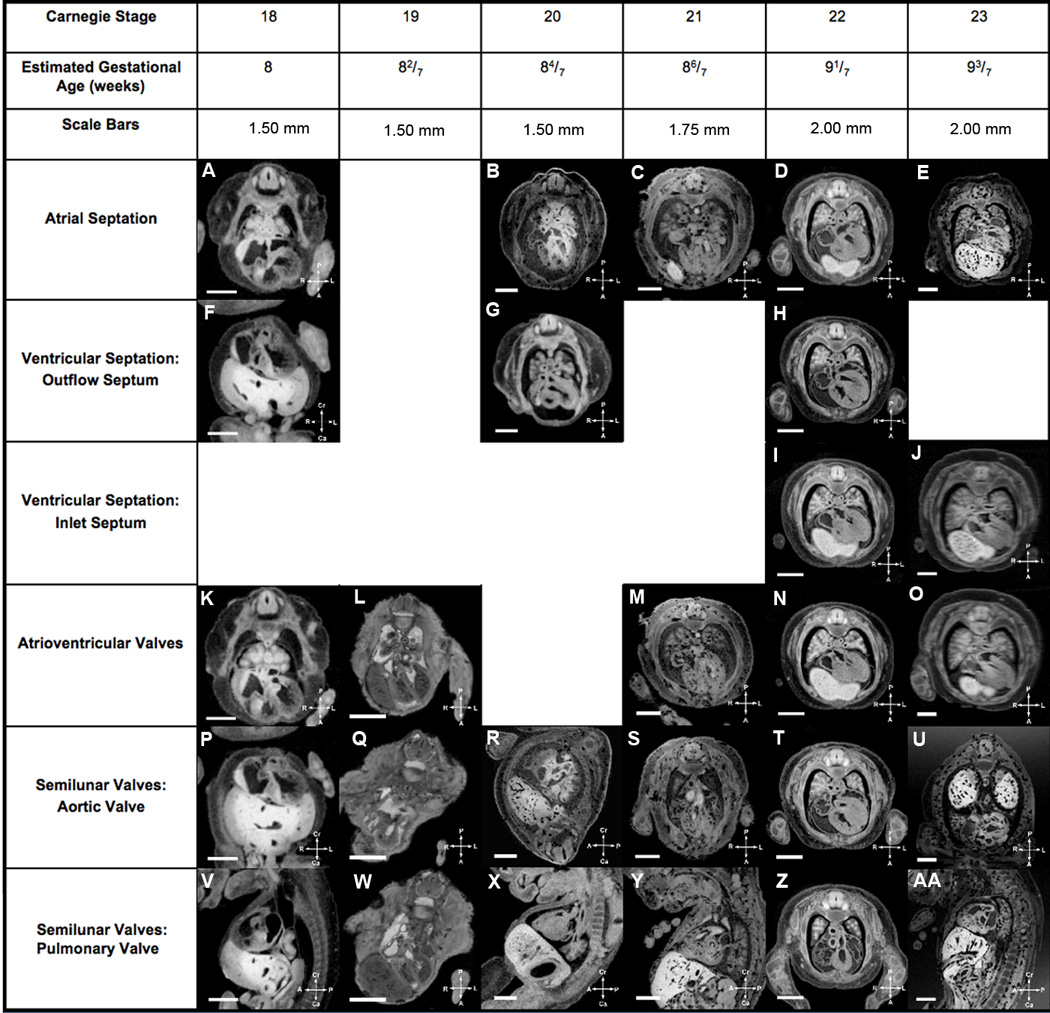
Major cardiac developmental structures present in EGA 6 4/7 weeks to 7 5/7 weeks (CS13–17) embryos are summarized in Figure 7, while Figure 8 shows the major developmental structures present in EGA 8 weeks to 9 3/7 weeks (CS18–23) embryos. One can look at a developmental structure at a specific estimated gestational age to determine what normal development is for that structure at that age in human cardiac development. The compass orients the observer to the plane of section.
References
-
- Kramer T. The partitioning of the truncus and conus and the formation of the membranous portion of the interventricular septum in the human heart. Am J Anat. 1942;71:343–370.
-
- Grant RP. The embryology of ventricular flow pathways in man. Circulation. 1962;25:756–779. - PubMed
-
- Goor DA, Edwards JE, Lillehei CW. The development of the interventricular septum of the human heart; correlative morphogenetic study. Chest. 1970;58:453–467. - PubMed
Publication types
MeSH terms
Grants and funding
LinkOut - more resources
Full Text Sources
Medical
