

MRI and CSF biomarkers in normal, MCI, and AD subjects: predicting future clinical change
- PMID: 19636049
- PMCID: PMC2715214
- DOI: 10.1212/WNL.0b013e3181af79fb
MRI and CSF biomarkers in normal, MCI, and AD subjects: predicting future clinical change
Abstract
Objective: To investigate the relationship between baseline MRI and CSF biomarkers and subsequent change in continuous measures of cognitive and functional abilities in cognitively normal (CN) subjects and patients with amnestic mild cognitive impairment (aMCI) and Alzheimer disease (AD) and to examine the ability of these biomarkers to predict time to conversion from aMCI to AD.
Methods: Data from the Alzheimer's Disease Neuroimaging Initiative, which consists of CN, aMCI, and AD cohorts with both CSF and MRI, were used. Baseline CSF (t-tau, Abeta(1-42), and p-tau(181P)) and MRI scans were obtained in 399 subjects (109 CN, 192 aMCI, 98 AD). Structural Abnormality Index (STAND) scores, which reflect the degree of AD-like features in MRI, were computed for each subject.
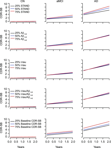
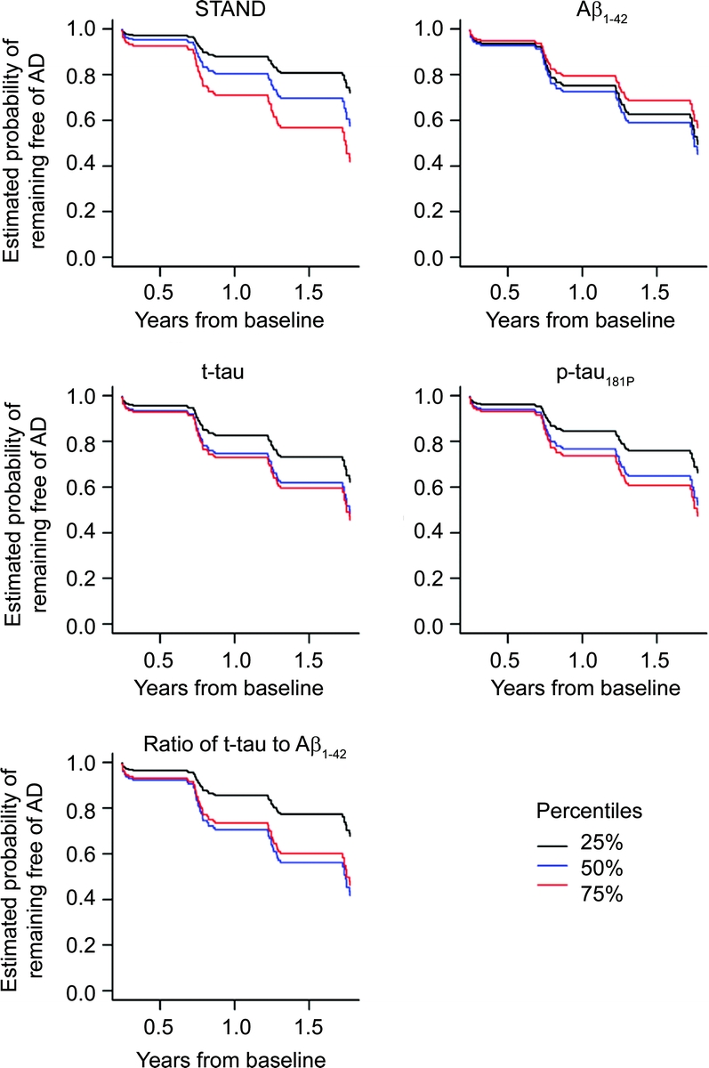
Results: Change on continuous measures of cognitive and functional performance was modeled as average Clinical Dementia Rating-sum of boxes and Mini-Mental State Examination scores over a 2-year period. STAND was a better predictor of subsequent cognitive/functional change than CSF biomarkers. Single-predictor Cox proportional hazard models for time to conversion from aMCI to AD showed that STAND and log (t-tau/Abeta(1-42)) were both predictive of future conversion. The age-adjusted hazard ratio for an interquartile change (95% confidence interval) of STAND was 2.6 (1.7, 4.2) and log (t-tau/Abeta(1-42)) was 2.0 (1.1, 3.4). Both MRI and CSF provided information about future cognitive change even after adjusting for baseline cognitive performance.
Conclusions: MRI and CSF provide complimentary predictive information about time to conversion from amnestic mild cognitive impairment to Alzheimer disease and combination of the 2 provides better prediction than either source alone. However, we found that MRI was a slightly better predictor of future clinical/functional decline than the CSF biomarkers tested.
Figures
References
-
- Price JL, Ko AI, Wade MJ, Tsou SK, McKeel DW, Morris JC. Neuron number in the entorhinal cortex and CA1 in preclinical Alzheimer disease. Arch Neurol 2001;58:1395–1402. - PubMed
-
- Tapiola T, Overmyer M, Lehtovirta M, et al. The level of cerebrospinal fluid tau correlates with neurofibrillary tangles in Alzheimer's disease. Neuroreport 1997;8:3961–3963. - PubMed
-
- Strozyk D, Blennow K, White LR, Launer LJ. CSF Abeta 42 levels correlate with amyloid-neuropathology in a population-based autopsy study. Neurology 2003;60:652–656. - PubMed
-
- Fagan AM, Roe CM, Xiong C, Mintun MA, Morris JC, Holtzman DM. Cerebrospinal fluid tau/beta-amyloid ratio as a prediction of cognitive decline in nondemented older adults. Arch Neurol 2007;64:343–349. - PubMed
Publication types
MeSH terms
Substances
Grants and funding
LinkOut - more resources
Full Text Sources
Other Literature Sources
Medical