. 2009 Oct 1;114(14):3008-17.
doi: 10.1182/blood-2008-04-148643. Epub 2009 Jul 27.

Role of leukemia cell invadosome in extramedullary infiltration

Affiliations

Role of leukemia cell invadosome in extramedullary infiltration

Michael Stefanidakis et al. Blood. .

Abstract

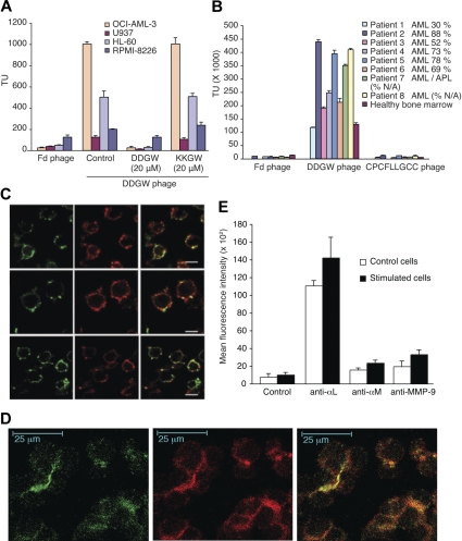
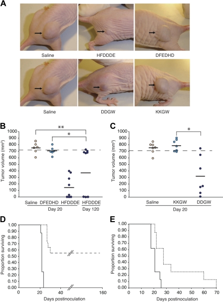
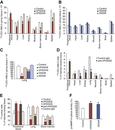
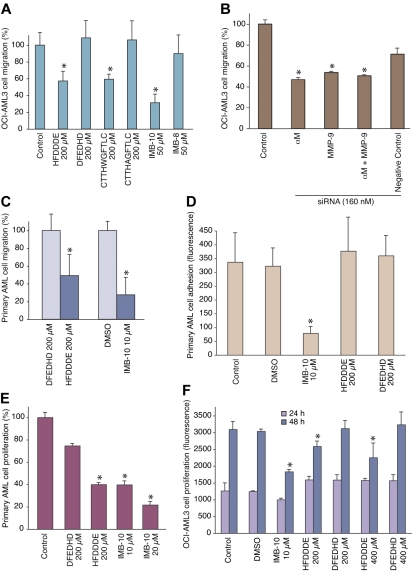
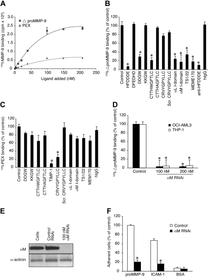
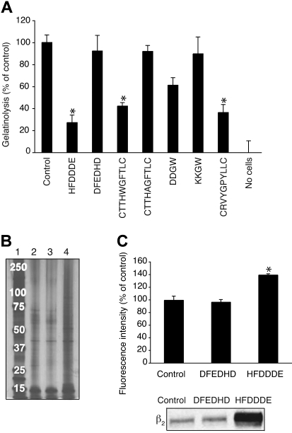
Acute myelogenous leukemias (AMLs) are characterized by medullary and extramedullary invasion. We hypothesized that a supramolecular complex, the leukemia-cell invadosome, which contains certain integrins, matrix metalloproteinases (MMPs), and other as-yet unidentified proteins, is essential for tissue invasion and may be central to the phenotypic diversity observed in the clinic. Here we show that the specific binding of MMP-9 to leukocyte surface beta(2) integrin is required for pericellular proteolysis and migration of AML-derived cells. An efficient antileukemia effect was obtained by the hexapeptide HFDDDE, a motif of the MMP-9 catalytic domain that mediates integrin binding: HFDDDE prevented proMMP-9 binding, transmigration through a human endothelial cell layer, and extracellular matrix degradation. Notably, the functional protein anchorage between beta(2) integrin and proMMP-9 described in this study does not involve the enzymatic active sites targeted by known MMP inhibitors. Taken together, our results provide a biochemical working definition for the human leukemia invadosome. Disruption of specific protein complexes within this supramolecular target complex may yield a new class of anti-AML drugs with anti-invasion (rather than or in addition to cytotoxic) attributes.


Figures

Figure 1
Figure 1
Expression of MMP-9 and integrin on AML cells and binding activity of DDGW peptide. (A) Phage displaying DDGW was allowed to bind to 4 leukemia cell lines in the absence or presence of DDGW or KKGW peptide. Insertless Fd phage was a control. The bound phages were determined by titering in bacteria. (B) Fd, DDGW, or CPCFLLGCC phages were examined for binding to bone marrow smears from AML patients and one healthy donor. The percentage of tumor cells is indicated. The bound phages were determined by bacterial infection. The results show mean ± SD. (C) OCI-AML3 cells were treated with 50 nmol/L PDBu for 30 minutes and stained with anti-αL TS1/22, anti-αM MEM170, and anti-β2 R7E4 antibodies (in green in top, middle, and bottom panels, respectively) and affinity purified antibodies against MMP-9 (in red). Yellow indicates the colocalization of αLMβ2 integrins and MMP-9. (D) OCI-AML3 cells were transferred onto the slides coated with endothelial cells (EaHy926), and the cells were treated with 50 nmol/L PDBu for 2 hours. Coculture was stained with anti-αM MEM170 (green) and polyclonal anti-MMP-9 H-129 (red) antibodies. Yellow indicates colocalization. (E) Cells were grown in the absence or presence of an endothelial cell monolayer and 50 nmol/L PDBu for 2 hours. The cells were stained with anti-αL MEM83, anti-αM MEM170, or anti-MMP-9 H-129 antibodies and analyzed by flow cytometry. Shown are mean ± SD from 3 separate experiments.
Figure 2
Figure 2
Antileukemia activity of invadosome-targeting peptides in OCI-AML3 xenografts. (A) Shown are representatives of peptide-treated leukemia-bearing mice 20 days after OCI-AML-3 cell inoculation. (B and C) Tumor sizes of OCI-AML-3–derived xenografts. Bars represent means from each peptide group. *Student t test, P < .001 of either HFDDDE- or DDGW-treated mice compared with control mice. No significant differences were detected between DFEDHD vs the saline group (t test, P = .107) or KKGW vs the saline group (t test, P = .65). Mice were killed when leukemia-derived xenograft volume reached 700 mm3 as indicated. (D-E) Kaplan-Meier actuarial survival analysis of the cohorts is shown. Differences were statistically significant at P < .001 and P = .004 for HFDDDE-treated group (D, dashed line) and DDGW-treated group (E, dashed line), respectively, compared with control DFEDHD-treated mice (D, solid line) or KKGW peptide-treated group (E, solid line).
Figure 3
Figure 3
Invadosome-targeting peptides reduce THP-1 xenograft growth and host cell infiltration. In THP-1–derived xenograft differences were statistically significant at P < .001 and P = .004 for HFDDDE- (A) or DDGW-treated groups (B), respectively, compared with vehicle-treated group. (C) Staining of tumor-infiltrating leukocytes with an αMβ2 integrin antibody (top panel) and tumor vasculature with an anti-CD31 monoclonal antibody (bottom panel). Representative tissue sections are shown from mice treated with saline, HFDDDE, or DDGW. Scale bar, 200 μm.
Figure 4
Figure 4
Effect of invadosome-inhibiting peptides on leukemia-cell extravasation and circulating MMP-9 levels. (A-D) 125I-labeled OCI-AML-3 cells were administered intravenously into Balb/c mice with peptide (200 μg), anti-HFDDDE (20 μg), or preimmune IgG. At 1 hour after inoculation, mice were killed, and tissues were harvested, weighed in, and subjected to a γ-counter. (E) 125I-labeled Jurkat leukemia T cells were administered as described previously. (F) Peptide alone was administered intravenously, and serum MMP-9 activity was measured by gelatin zymography. (A-F) Shown are means ± SD from triplicates. *P < .005 (t test) of HFDDDE or antibody-treated mice compared with control mice.
Figure 5
Figure 5
Inhibition of AML cell migration, adhesion, and proliferation in vitro. (A) OCI-AML3 cells treated with 200 μmol/L peptide or 20 μmol/L small-molecule were allowed to migrate through an endothelial-cell monolayer. The results show means ± SD from triplicate wells. *P < .001 by Student t test. (B) Cells pretreated with RNAi oligomers were assessed as in panel A; *P < .001. (C) AML-M4 primary human leukemia-derived cells were subjected to migration in collagen-coated chambers for 24 hours, and the migrated cells were counted. DMSO indicates dimethylsulfoxide. *P < .05. (D) AML-M4 primary human leukemia-derived cells were allowed to bind to gelatin-coated microtiter wells for 60 minutes after which the bound cells were determined via the DHL assay; *P < .001. (E) AML-M4 primary cells were cultivated in suspension for 7 days with the compounds as described and the growth was determined via the DHL assay; *P < .02. (F) OCI-AML3 cells were cultivated in suspension for 24 hours or 48 hours, and the growth was determined via the DHL assay; *P < .01.
Figure 6
Figure 6
The integrin I-domain anchors proMMP-9 at the cell surface. (A) 125I-labeled MMP-9 domains bind to OCI-AML-3 cells in a dose-dependent manner. (B) 125I-ΔproMMP-9 binding was competed with peptides (200 μmol/L), αL and αM I domains (20 μg/mL), or the antibodies (20 μg). (C) Binding of 125I-PEX domain was studied as in panel B. (D) 125I-ΔproMMP-9 binding to siRNA-treated or untreated cells. (E) Immunoblots of siRNA-treated cells with αM antibodies and control α-actinin antibodies. (F) Binding of siRNA-treated cells to immobilized proMMP-9, intercellular adhesion molecule-1, or BSA. Adherent cells were quantitated by phosphatase assay. Data are means ± SD from triplicates. *P < .001 by t test.
Figure 7
Figure 7
Inhibition of pericellular proteolysis of OCI-AML3 cells. (A) Effects of peptides (200 μmol/L) on the release of gelatin fragments from coated FITC-labeled gelatin. The results show means ± SD from triplicates. *P < .005. (B) Biotinylated cell surface proteins were isolated from cells incubated without peptide (lane 2), with DFEDHD (lane 3), or with HFDDDE (lane 4). Samples were run on sodium dodecyl sulfate–polyacrylamide gel electrophoresis followed by silver staining. Molecular weight markers are shown in lane 1. (C) Quantitation of cell-surface biotinylated proteins with streptavidin-phycoerythrinin flow cytometry (top panel). Immunoblotting with β2 integrin antibody (bottom panel).

Similar articles

Cited by

References

    1. Scheinberg D, Maslak P, Weiss M. In: Cancer: Principles and Practice of Oncology. Vita V, Hellman S, Rosenberg S, editors. Philadelphia, PA: Lippincott-Raven; 2005. pp. 2404–2432.
    1. Jandl JH. Blood: Textbook of Haematology. 2nd ed. Toronto: Little, Brown and Co; 1996. pp. 1–1200.
    1. Stefanidakis M, Koivunen E. Cell-surface association between matrix metalloproteinases and integrins: role of the complexes in leukocyte migration and cancer progression. Blood. 2006;108(5):1441–1450. - PubMed
    1. Björklund M, Aitio O, Stefanidakis M, et al. Stabilization of the activated aMb2 integrin by a small-molecule inhibits leukocyte migration and recruitment. Biochemistry. 2006;45(9):2862–2871. - PubMed
    1. Suojanen J, Salo T, Sorsa T, Koivunen E. aMb2 integrin modulator exerts antitumor activity in vivo. Anticancer Res. 2007;27(6B):3775–3782. - PubMed

Publication types

MeSH terms