

doi: 10.1088/0031-9155/54/16/N01.
Epub 2009 Jul 27.
Optical imaging of Cerenkov light generation from positron-emitting radiotracers
Affiliations
- PMID: 19636082
- PMCID: PMC2765256
- DOI: 10.1088/0031-9155/54/16/N01
Item in Clipboard
Optical imaging of Cerenkov light generation from positron-emitting radiotracers
Phys Med Biol.
.
Abstract
Radiotracers labeled with high-energy positron emitters, such as those commonly used for positron emission tomography studies, emit visible light immediately following decay in a medium. This phenomenon, not previously described for these imaging tracers, is consistent with Cerenkov radiation and has several potential applications, especially for in vivo molecular imaging studies. Herein we detail a new molecular imaging tool, Cerenkov Luminescence Imaging, the experiments conducted that support our interpretation of the source of the signal, and proof-of-concept in vivo studies that set the foundation for future application of this new method.
Figures
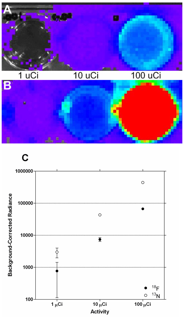
The luminescence detected from well plates of 1, 10 and 100 µCi of 18F (A) or 13N (B) following a 10 second image acquisition. The quantification of the radiance, plotted in C, revealed that the higher energy positron-emitter produced significantly more light. For example, at 100 µCi, 13N produces approximately 6.5× more light than 18F.
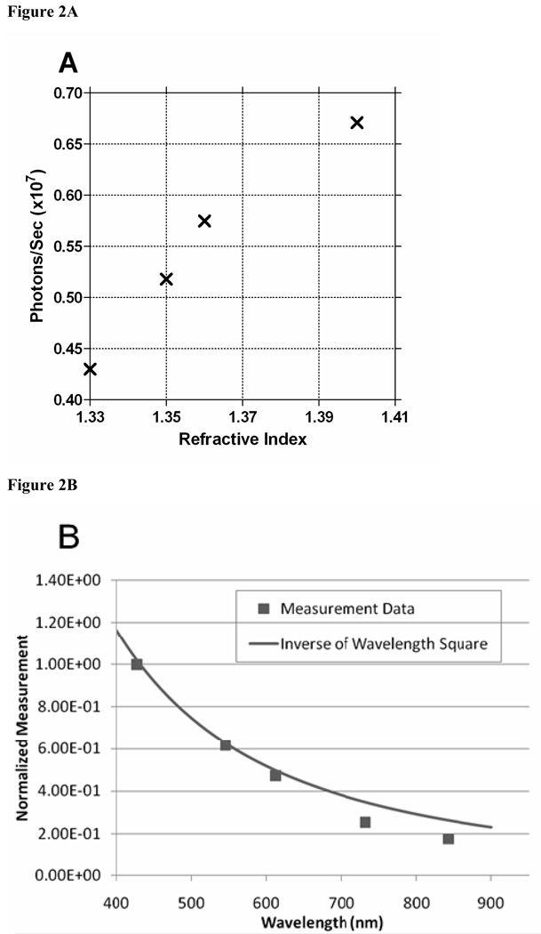
A: The light output from solutions of solutions with refractive indices of approximately 1.33, 1.35, 1.37, and 1.41 (0, 10, 20, and 40% glucose) spiked with 209 µCi FDG. The light output increase as a function of glucose concentration (increase in refractive index) is consistent with the reduction in the speed of the propagation of light in the medium and an amplification of the Cerenkov radiation. B: Results from a series of filtered scans on the wavelength of light detected from positron-emitter samples. The majority of the light is produced in the blue portion of the spectrum, which is expected for Cerenkov radiation, and follows an inverse relationship with the square of the wavelength. Cerenkov light extends into the green and red region of the EM spectrum, which could increase the range of applications for in vivo imaging studies.
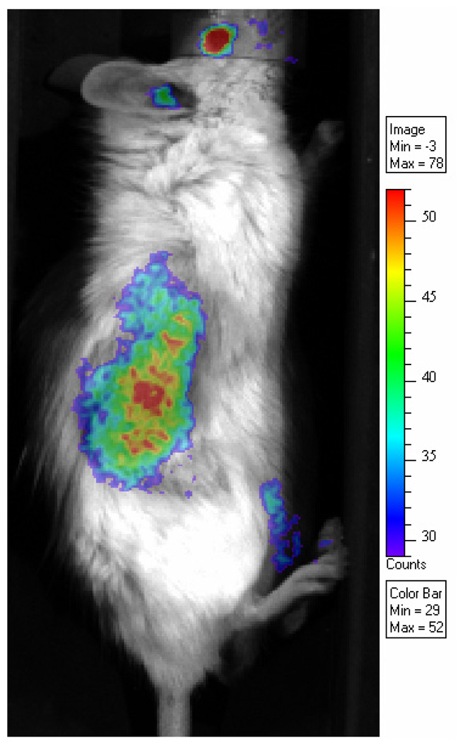
Optical scan of a mouse bearing a CWR22-RV1 xenograft following injection of 270 µCi FDG. Luminescence was detected throughout the animal, which is consistent with the broad distribution of FDG; however, for display, the image is thresholded to highlight the tumor region. In the tumor, the measured luminescence is 11× the signal measured in a region above the tumor and 47× the signal from the image background.
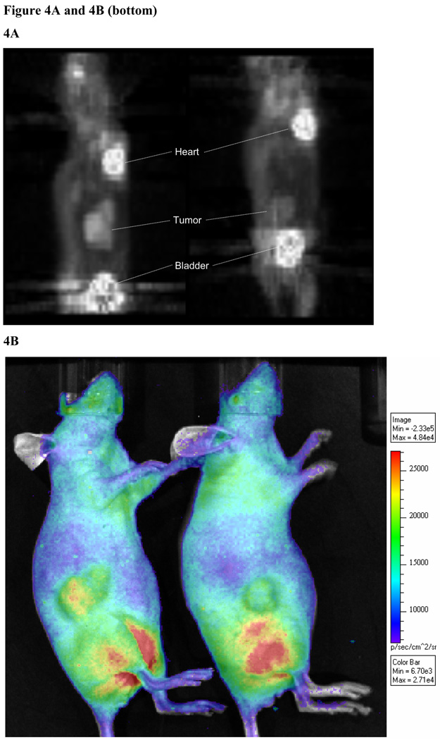
A sagittal maximum intensity projection (MIP) of an FDG PET scan on two mice bearing flank colon tumors derived from primary human tissue (A). The image were injected with 300 µCi FDG and prior to the PET scan (10 min) were imaged in the optical scanner (1 min exposure). The optical scan (Figure 4B) showed different degrees of light output that were consistent with the quantification from the PET scan. The SUV was 2.4 and 1.9, and the %ID/g was 9.8% and 7.4% (left and right, respectively). From the optical image, a similar intensity difference was measured: 4.4×10⁵ photons/s versus 2.3×10⁵ photons/s, left and right, respectively. In the optical scan, the image was thresholded to show the light emanating from the animal, demonstrating the full distribution of the FDG tracer as shown in PET scans.
Similar articles
-
Cerenkov imaging.Adv Cancer Res. 2014;124:213-34. doi: 10.1016/B978-0-12-411638-2.00006-9. Adv Cancer Res. 2014. PMID: 25287690 Free PMC article. Review.
-
Cerenkov luminescence tomography for small-animal imaging.Opt Lett. 2010 Apr 1;35(7):1109-11. doi: 10.1364/OL.35.001109. Opt Lett. 2010. PMID: 20364233 Free PMC article.
-
Cerenkov luminescence imaging of medical isotopes.J Nucl Med. 2010 Jul;51(7):1123-30. doi: 10.2967/jnumed.110.076521. Epub 2010 Jun 16. J Nucl Med. 2010. PMID: 20554722 Free PMC article.
-
Comments on 'Cerenkov radiation allows in vivo optical imaging of positron emitting radiotracers'.Phys Med Biol. 2010 Sep 21;55(18):L43-4; author reply L45-9. doi: 10.1088/0031-9155/55/18/L01. Epub 2010 Aug 24. Phys Med Biol. 2010. PMID: 20736495
-
Optical imaging as an expansion of nuclear medicine: Cerenkov-based luminescence vs fluorescence-based luminescence.Eur J Nucl Med Mol Imaging. 2013 Aug;40(8):1283-91. doi: 10.1007/s00259-013-2408-9. Epub 2013 May 15. Eur J Nucl Med Mol Imaging. 2013. PMID: 23674205 Review.
Cited by
-
Cerenkov luminescence imaging is an effective preclinical tool for assessing colorectal cancer PD-L1 levels in vivo.EJNMMI Res. 2020 Jun 15;10(1):64. doi: 10.1186/s13550-020-00654-w. EJNMMI Res. 2020. PMID: 32542442 Free PMC article.
-
Imaging Cherenkov photon emissions in radiotherapy with a Geiger-mode gated quanta image sensor.Opt Lett. 2019 Sep 15;44(18):4546-4549. doi: 10.1364/OL.44.004546. Opt Lett. 2019. PMID: 31517927 Free PMC article.
-
Signal intensity analysis and optimization for in vivo imaging of Cherenkov and excited luminescence.Phys Med Biol. 2018 Apr 20;63(8):085019. doi: 10.1088/1361-6560/aab83b. Phys Med Biol. 2018. PMID: 29558363 Free PMC article.
-
PEGylated crushed gold shell-radiolabeled core nanoballs for in vivo tumor imaging with dual positron emission tomography and Cerenkov luminescent imaging.J Nanobiotechnology. 2018 Apr 18;16(1):41. doi: 10.1186/s12951-018-0366-x. J Nanobiotechnology. 2018. PMID: 29669544 Free PMC article.
-
Anti-CD45 radioimmunotherapy with 90Y but not 177Lu is effective treatment in a syngeneic murine leukemia model.PLoS One. 2014 Dec 2;9(12):e113601. doi: 10.1371/journal.pone.0113601. eCollection 2014. PLoS One. 2014. PMID: 25460570 Free PMC article.
References
-
- Clarke LP, Cullom SJ, Shaw R, Reece C, Penney BC, King MA, Silbiger M. Bremsstrahlung imaging using the gamma camera: factors affecting attenuation. J. Nucl. Med. 1992;33:161–166. - PubMed
-
- Collins GB, Reiling VG. Cerenkov Radiation. Phys. Rev. 1938;54:499–503.
-
- Elrick RH, Parker RP. The use of Cerenkov radiation in the measurement of beta-emitting radionuclides. Int. J. Appl. Radiat. Isot. 1968;19:263–271. - PubMed
-
- Jelley JV. Cerenkov radiation and its applications. Br. J. Appl. Phys. 1955;6:227–232.
-
- Kelloff GJ, Hoffman JM, Johnson B, Scher HI, Siegel BA, Cheng EY, Cheson BD, O'Shaughnessy J, Guyton KZ, Mankoff MA, Shankar L, Larson SM, Sigman CC, Schilsky RL, Sullivan DC. Progress and Promise of FDG-PET Imaging for Cancer Patient Management and Oncologic Drug Development. Clin. Cancer Res. 2005;11:2785–2808. - PubMed
MeSH terms
Substances
Grants and funding
LinkOut - more resources
Full Text Sources
Other Literature Sources