. 2009 Aug;31(8):561-70.
doi: 10.1097/MPH.0b013e3181a6e043.

Therapeutic vulnerability of an in vivo model of alveolar soft part sarcoma (ASPS) to antiangiogenic therapy

Affiliations

Therapeutic vulnerability of an in vivo model of alveolar soft part sarcoma (ASPS) to antiangiogenic therapy

David T Vistica et al. J Pediatr Hematol Oncol. 2009 Aug.

Abstract

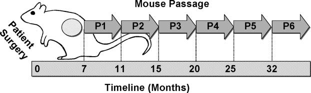
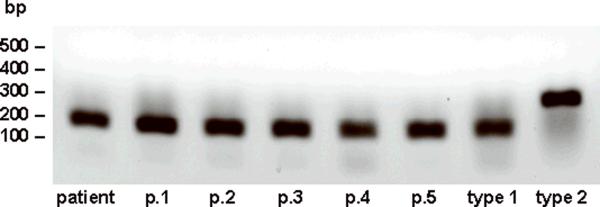
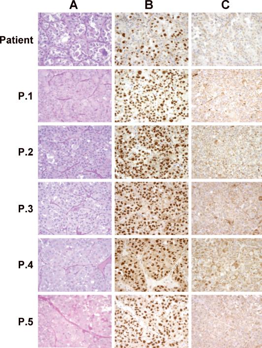
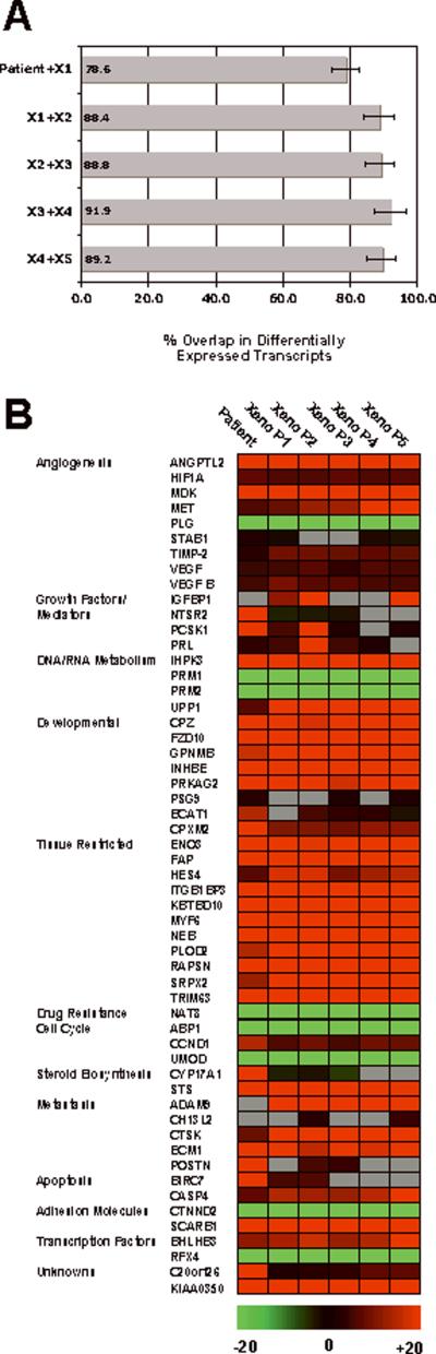
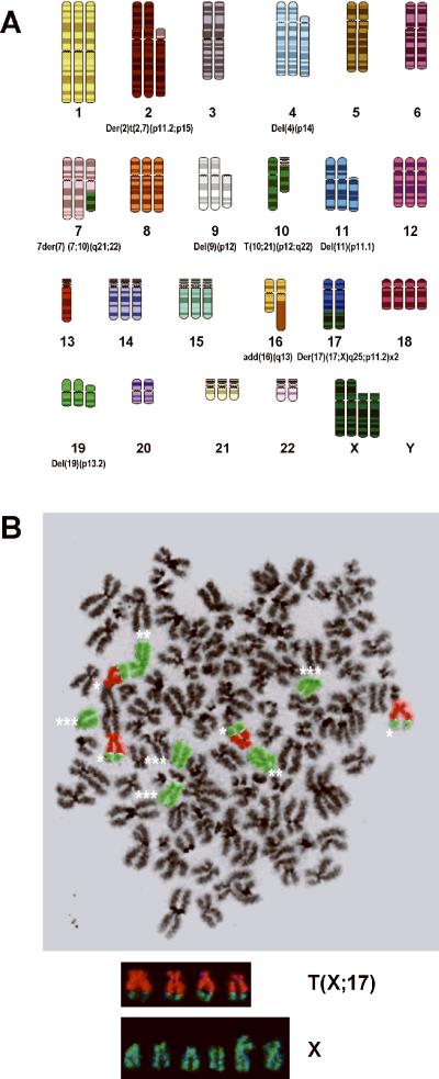
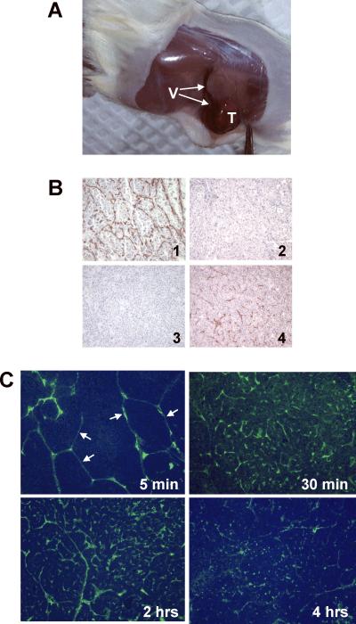
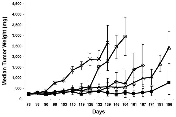
In vivo growth of alveolar soft part sarcoma (ASPS) was achieved using subcutaneous xenografts in sex-matched nonobese diabetic severe combined immunodeficiency mice. One tumor, currently at passage 6, has been maintained in vivo for 32 months and has maintained characteristics consistent with those of the original ASPS tumor including (1) tumor histology and staining with periodic acid Schiff/diastase, (2) the presence of the ASPL-TFE3 type 1 fusion transcript, (3) nuclear staining with antibodies to the ASPL-TFE3 type 1 fusion protein, (4) maintenance of the t(X;17)(p11;q25) translocation characteristic of ASPS, (5) stable expression of signature ASPS gene transcripts and finally, the development and maintenance of a functional vascular network, a hallmark of ASPS. The ASPS xenograft tumor vasculature encompassing nests of ASPS cells is highly reactive to antibodies against the endothelial antigen CD34 and is readily accessible to intravenously administered fluorescein isothiocyanate-dextran. The therapeutic vulnerability of this tumor model to antiangiogenic therapy, targeting vascular endothelial growth factor and hypoxia-inducible factor-1 alpha, was examined using bevacizumab and topotecan alone and in combination. Together, the 2 drugs produced a 70% growth delay accompanied by a 0.7 net log cell kill that was superior to the antitumor effect produced by either drug alone. In summary, this study describes a preclinical in vivo model for ASPS which will facilitate investigation into the biology of this slow growing soft tissue sarcoma and demonstrates the feasibility of using an antiangiogenic approach in the treatment of ASPS.


Figures

Figure 1
Figure 1
Growth and Serial Passage of ASPS Xenograft Tumors in Immunocompromised NOD.SCID\NCr Mice. ASPS tumor fragments (1-2 mm) were implanted subcutaneously in NOD.SCID\NCr immunocompromised mice within 24 hours following patient surgery as described in Materials and Methods. Tumors were harvested at the indicated times and serially passaged.
Figure 2
Figure 2
RT-PCR Determination of the ASPL-TFE3 Fusion Transcript. RNA was isolated from ASPS xenograft tumors at the appropriate passage and the ASPL-TFE3 fusion transcript was determined by RT-PCR as described in Materials and Methods.
Figure 3
Figure 3
ASPS Xenograft Histology and Nuclear Reactivity to ASPL-TFE3 type 1 and ASPL-TFE3 type 2 Antibodies. ASPS xenograft tumors, maintained in female NOD.SCID\NCr mice, were harvested at the indicated passage (see timeline illustrated in Figure 1), fixed in 10% buffered formalin and embedded in paraffin. Sections (4μm) were stained with Periodic Acid Schiff (PAS)( Panel A) or with affinity-purified polyclonal antibodies to either the ASPLTFE3 type 1 fusion protein (Panel B) or to the ASPL-TFE3 type 2 fusion protein (Panel C). (See reference 3 for details of staining).
Figure 4
Figure 4
Conservation and Stability of Selected Genes in ASPS Xenograft Tumors. Panel A. Transcript Conservation in ASPS Xenograft Tumors. Data was analyzed as described in Materials and Methods. Panel B. ASPS Xenograft Tumor Heat Map. Data was analyzed as described in Materials and Methods.
Figure 5
Figure 5
ASPS Xenograft Tumors exhibit a Near-Triploid Karyotype and the t(X;17)(p11;q25) Chromosomal Translocation. Panel A. ASPS Xenograft Spectral Karyotype (SKY). Numerous chromosomal aberrations are present in this near triploid karyotype including the characteristic t(X;17)(q11;p25) translocation. Panel B. FISH Analysis of the ASPS Xenograft Tumor. Four copies of the t(X;17)(q11;p25) translocation (*), 2 normal X chromosomes (**) and 4 deleted X chromosomes (***) are present. X chromosome (green); chromosome 17 (orange).
Figure 6
Figure 6
ASPS Xenograft Tumor Vasculature. Panel A. ASPS Xenograft Tumors Exhibit a Complex Vascular Network. Numerous blood vessels (V) can be seen entering the subcutaneous ASPS tumor (T). Panel B. CD31 and CD34 Staining of the ASPS Endothelial Network. ASPS xenograft tumors were harvested, fixed in 10% buffered formalin, embedded in paraffin and stained (3) with antibodies to human and mouse CD34. Panel 1: Patient Tumor (anti-human CD34). Panel 2: Patient Tumor (anti-mouse CD34). Panel 3: ASPS Xenograft Tumor (anti-human CD 34). Panel 4: ASPS Xenograft Tumor (anti-mouse CD 34). Panel C. FITC-Dextran Localizes to the Endothelium Surrounding Nests of ASPS Cells Following Intravenous Administration. ASPS tumors were harvested at 5, 30, 120 and 240 minutes following intravenous administration of the fluorochrome and processed as described in Material and Methods. The fluorochrome readily locates to the endothelial network surrounding nests of ASPS tumor cells (5 minutes; arrows) and slowly diffuses into the tumor.
Figure 7
Figure 7
Antiangiogenic Therapy of ASPS Xenograft Tumors. Median tumor weights of mice treated with bevacizumab, topotecan or a combination of both drugs. Female NOD.SCID mice bearing subcutaneous ASPS tumors were randomized into one of 5 groups (n=9/group): ---x--- vehicle control Q3D x 6; ---□ ---100 ug bevacizumab Q3D x 6; ---◊--- 12 mg topotecan/kg Q4D x 4; ---Δ---1mg topotecan/kg QD x 15; ---■--- 100ug bevacizumab Q3D x 6 and 1 mg topotecan/kg QD x 15. All treatments were administered intraperitoneally starting on day 76 when tumors staged approximately 230mg. Two additional treatment cycles were initiated on days 96 and 116. Mice were euthanized when tumors reached a size of 2500mg or at the termination of the experiment.

References

    1. Christopherson WM, Foote FW, Jr, Stewart FW. Alveolar soft-part sarcomas: structurally characteristic tumors of uncertain histogenesis. Cancer. 1952;5(1):100–111. - PubMed
    1. Shipkey FH, Lieberman PH, Foote FW, Jr., et al. Ultrastructure of alveolar soft part sarcoma. Cancer. 1964;17:821–830. - PubMed
    1. Vistica DT, Krosky PM, Kenney S, et al. Immunohistochemical discrimination between the ASPL-TFE3 fusion proteins of alveolar soft part sarcoma. J Pediatr Hematol Oncol. 2008;30:46–52. - PubMed
    1. Wu Z, Irizarry RA. Stochastic models inspired by hybridization theory for short oligonucleotide arrays. J Comput Biol. 2005;12(6):882–893. - PubMed
    1. Seabright M. A rapid banding technique for human chromosomes. Lancet. 1971;11:971–972. - PubMed

Publication types

MeSH terms

Substances