

Linking white and grey matter in schizophrenia: oligodendrocyte and neuron pathology in the prefrontal cortex
- PMID: 19636386
- PMCID: PMC2713751
- DOI: 10.3389/neuro.05.009.2009
Linking white and grey matter in schizophrenia: oligodendrocyte and neuron pathology in the prefrontal cortex
Abstract
Neuronal circuitry relies to a large extent on the presence of functional myelin produced in the brain by oligodendrocytes. Schizophrenia has been proposed to arise partly from altered brain connectivity. Brain imaging and neuropathologic studies have revealed changes in white matter and reduction in myelin content in patients with schizophrenia. In particular, alterations in the directionality and alignment of axons have been documented in schizophrenia. Moreover, the expression levels of several myelin-related genes are decreased in postmortem brains obtained from patients with schizophrenia. These findings have led to the formulation of the oligodendrocyte/myelin dysfunction hypothesis of schizophrenia. In this review, we present a brief overview of the neuropathologic findings obtained on white matter and oligodendrocyte status observed in schizophrenia patients, and relate these changes to the processes of brain maturation and myelination. We also review recent data on oligodendrocyte/myelin genes, and present some recent mouse models of myelin deficiencies. The use of transgenic and mutant animal models offers a unique opportunity to analyze oligodendrocyte and neuronal changes that may have a clinical impact. Lastly, we present some recent morphological findings supporting possible causal involvement of white and grey matter abnormalities, in the aim of determining the morphologic characteristics of the circuits whose alteration leads to the cortical dysfunction that possibly underlies the pathogenesis of schizophrenia.
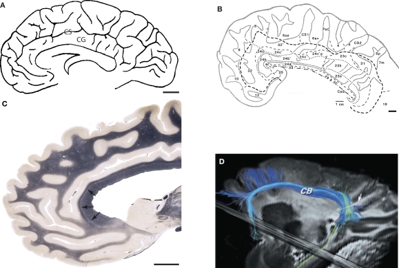
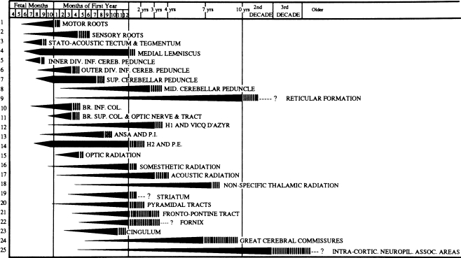
Keywords: anterior cingulate cortex; cingulum bundle; development; myelin; myelin-related genes.
Figures
Similar articles
-
Glutathione deficit impairs myelin maturation: relevance for white matter integrity in schizophrenia patients.Mol Psychiatry. 2015 Jul;20(7):827-38. doi: 10.1038/mp.2014.88. Epub 2014 Aug 26. Mol Psychiatry. 2015. PMID: 25155877
-
Spatial distribution and density of oligodendrocytes in the cingulum bundle are unaltered in schizophrenia.Acta Neuropathol. 2009 Apr;117(4):385-94. doi: 10.1007/s00401-008-0379-x. Epub 2008 Apr 26. Acta Neuropathol. 2009. PMID: 18438678 Free PMC article.
-
Is psychosis a dysmyelination-related information-processing disorder?Psychiatriki. 2019 Jul-Sep;30(3):245-255. doi: 10.22365/jpsych.2019.303.245. Psychiatriki. 2019. PMID: 31685456 Review.
-
Myelin and oligodendrocyte lineage cells in white matter pathology and plasticity after traumatic brain injury.Neuropharmacology. 2016 Nov;110(Pt B):654-659. doi: 10.1016/j.neuropharm.2015.04.029. Epub 2015 May 9. Neuropharmacology. 2016. PMID: 25963414 Review.
-
Oligodendrocyte morphometry and expression of myelin - Related mRNA in ventral prefrontal white matter in major depressive disorder.J Psychiatr Res. 2015 Jun;65:53-62. doi: 10.1016/j.jpsychires.2015.04.010. Epub 2015 Apr 20. J Psychiatr Res. 2015. PMID: 25930075 Free PMC article.
Cited by
-
Gray matter myelination of 1555 human brains using partial volume corrected MRI images.Neuroimage. 2015 Jan 15;105:473-85. doi: 10.1016/j.neuroimage.2014.10.054. Epub 2014 Nov 1. Neuroimage. 2015. PMID: 25449739 Free PMC article.
-
Increased density of DISC1-immunoreactive oligodendroglial cells in fronto-parietal white matter of patients with paranoid schizophrenia.Eur Arch Psychiatry Clin Neurosci. 2016 Sep;266(6):495-504. doi: 10.1007/s00406-015-0640-y. Epub 2015 Aug 28. Eur Arch Psychiatry Clin Neurosci. 2016. PMID: 26315603
-
Sequence of Molecular Events during the Maturation of the Developing Mouse Prefrontal Cortex.Mol Neuropsychiatry. 2015 Jul;1(2):94-104. doi: 10.1159/000430095. Epub 2015 Jun 9. Mol Neuropsychiatry. 2015. PMID: 26457295 Free PMC article.
-
Editorial: Neuroepigenetics of Neuropsychiatric Disease-Hope, Success and Obstacles for Translational Findings and Applications.Front Neurosci. 2022 Apr 1;16:886695. doi: 10.3389/fnins.2022.886695. eCollection 2022. Front Neurosci. 2022. PMID: 35431770 Free PMC article. No abstract available.
-
White matter abnormalities and animal models examining a putative role of altered white matter in schizophrenia.Schizophr Res Treatment. 2011;2011:826976. doi: 10.1155/2011/826976. Epub 2011 Aug 11. Schizophr Res Treatment. 2011. PMID: 22937274 Free PMC article.
References
-
- Akbarian S., Kim J. J., Potkin S. G., Hetrick W. P., Bunney W. E., Jr, Jones E. G. (1996). Maldistribution of interstitial neurons in prefrontal white matter of the brains of schizophrenic patients. Arch. Gen. Psychiatry 53, 425–436 - PubMed
Grants and funding
LinkOut - more resources
Full Text Sources