. 2009 Aug 21;9(16):2339-47.
doi: 10.1039/b907860f. Epub 2009 Jun 3.

Dynamics of individual polymers using microfluidic based microcurvilinear flow

Affiliations

Dynamics of individual polymers using microfluidic based microcurvilinear flow

Chao-Min Cheng et al. Lab Chip. .

Abstract

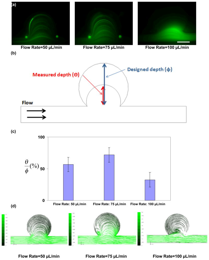
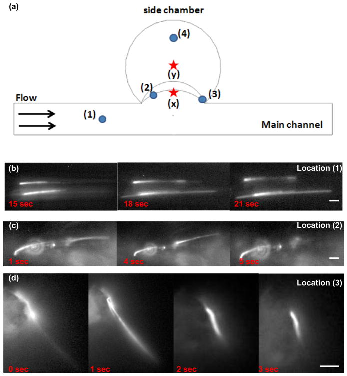
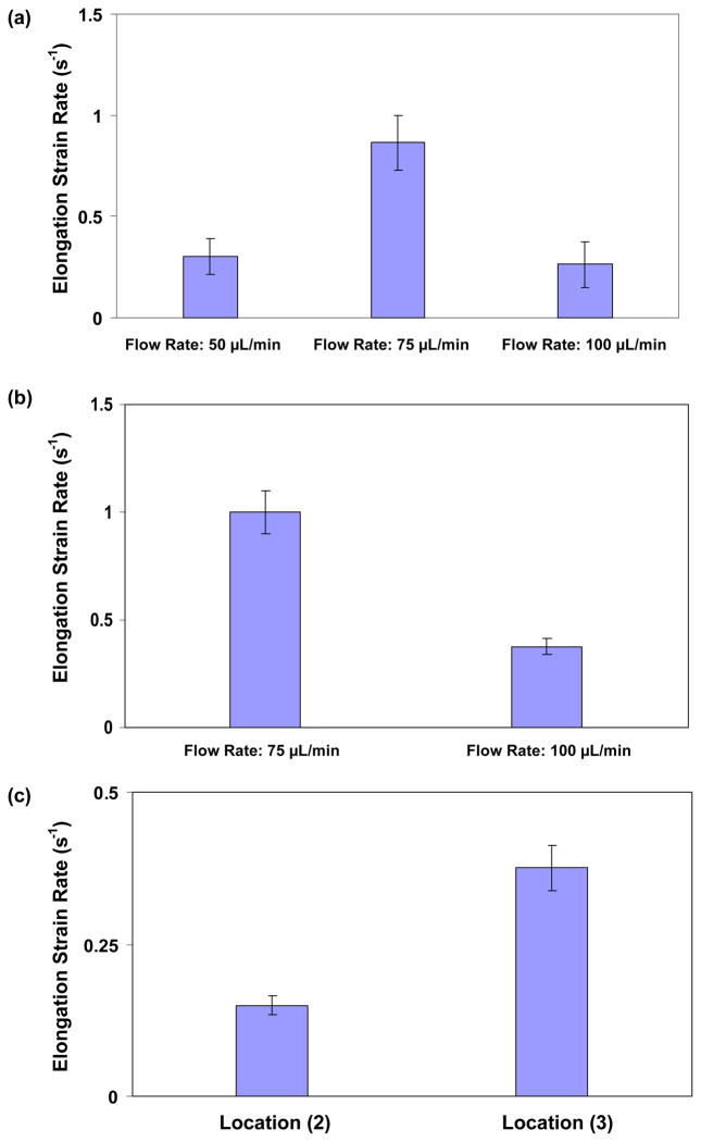
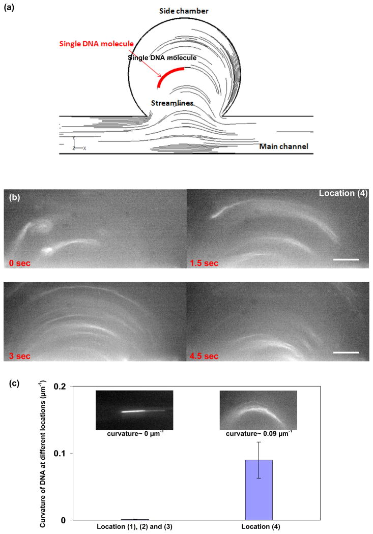
Polymer dynamics play an important role in a diversity of fields including materials science, physics, biology and medicine. The spatiotemporal responses of individual molecules such as biopolymers have been critical to the development of new materials, the expanded understanding of cell structures including cytoskeletal dynamics, and DNA replication. The ability to probe single molecule dynamics however is often limited by the availability of small-scale technologies that can manipulate these systems to uncover highly intricate behaviors. Advances in micro- and nano-scale technologies have simultaneously provided us with valuable tools that can interface with these systems including methods such as microfluidics. Here, we report on the creation of micro-curvilinear flow through a small-scale fluidic approach, which we have been used to impose a flow-based high radial acceleration ( approximately 10(3) g) on individual flexible polymers. We were able to employ this microfluidic-based approach to adjust and control flow velocity and acceleration to observe real-time dynamics of fluorescently labeled lambda-phage DNA molecules in our device. This allowed us to impose mechanical stimulation including stretching and bending on single molecules in localized regimes through a simple and straightforward technology-based method. We found that the flexible DNA molecules exhibited multimodal responses including distinct conformations and controllable curvatures; these characteristics were directly related to both the elongation and bending dynamics dictated by their locations within the curvilinear flow. We analyzed the dynamics of these individual molecules to determine their elongation strain rates and curvatures ( approximately 0.09 microm(-1)) at different locations in this system to probe the individual polymer structural response. These results demonstrate our ability to create high radial acceleration flow and observe real-time dynamic responses applied directly to individual DNA molecules. This approach may also be useful for studying other biologically based polymers including additional nucleic acids, actin filaments, and microtubules and provide a platform to understand the material properties of flexible polymers at a small scale.


Figures

Fig. 1
Fig. 1
Pressure-driven micro curvilinear flow design for application of high radial acceleration on individual DNA molecules. (a) Schematic of the pressure-driven fluidic system designed to observe the response dynamics of individual DNA molecules. The microfluidic device was fabricated through soft lithography and the flow rate was controlled via a syringe pump. (b) Diagram of the microfluidic configuration used to create the micro curvilinear flow. The surface of the glass substrate at the bottom of this micro curvilinear flow system was treated with anti-digoxigenin for at least one hour to attach the DNA molecules to the surface of the glass. (c) An optical image of the poly(dimethylsiloxane) microfluidic device along with, (d) a differential interference contrast image of the circular side chamber. Scale bar= 30 μm. (e) Fluorescent image of a YOYO-1 labeled DNA molecule within the channel of this micro curvilinear flow system. Scale bar=4 μm.
Fig. 2
Fig. 2
Effect of velocity on flow profile for the circular side chamber of the micro curvilinear flow device. (a) Fluorescent images of beads moving from the main channel into the side chamber under flow rates of 50 μL/min, 75 μL/min, and 100 μL/min. At flow rates that were higher and lower than these, the formation of a vortex flow pattern was inhibited. The rotational velocity and radial acceleration at the inner regime of this side chamber were 6.2 and 4.6 times higher than those observed at the outer regime. Scale bar= 30 μm. (b) Schematic of the micro curvilinear flow flow depth penetration compared with the designed vortex depth (the size of the side chamber). (c) Percentage of the average depth of penetration or distance into the side chamber when compared to the entire distance across the entire circular chamber. The flow rates were compared to the bead flow profile captured from these increased time exposure images; this was used to characterize the flow profile based on the velocity of the fluid (error bars represent the standard deviation). (d) Streamlines with green scale velocity magnitudes from the computational fluid dynamics simulation in the micro curvilinear flow system at different flow rates.
Fig. 3
Fig. 3
Spatial locations within the micro curvilinear flow to examine DNA response. (a) Schematic of specific spatial locations of the DNA molecules within the micro curvilinear flow side chamber. (b) Time-lapse snapshots of two individual DNA molecules in the main channel under a pressure-driven flow at a flow rate of 50 μL/min at location (1). Before introducing the DNA, we treated the surface of the glass substrate with anti-digoxigenin for at least one hour to induce the attachment of the DNA to the surface. Scale bar= 2 μm. (c) Time-lapse snapshots of an individual DNA molecule at location (2) in the micro curvilinear flow at a flow rate of 100 μL/min. Scale bar= 2 μm. (d) Time-lapse snapshots of an individual DNA molecule at location (3) under a flow rate of 75 μL/min. Scale bar= 4 μm. We measured the change in DNA length between two images with ImageJ (a public software from National Institutes of Health; http://rsbweb.nih.gov/ij/). Through using the elapsed time, we estimated the elongation strain and elongation strain rate of the DNA molecules.
Fig. 4
Fig. 4
(a) Elongation strain rates versus flow rates within the micro curvilinear flow system (error bars represent the standard deviation; 24 images from 5 samples). (b) Elongation strain rates versus flow rates at location (3) in Figure 3(a). The elongation strain rate at a flow rate of 75 μL/min is 2.66 times higher than for 100 μL/min (error bars represent the standard deviation). (c) Elongation strain rates versus a constant flow rate of 100 μL/min at different locations (location (2) and (3) in Figure 3(a)). The elongation strain rate at location (3) is 2.51 times greater than at location (2) (error bars represent the standard deviation).
Fig. 5
Fig. 5
(a) Schematic of conformation of a single DNA molecule at location (4) in the micro curvilinear flow. (b) Conformation of single DNA molecules in the micro curvilinear flow system under high radial acceleration. Fluorescent images of individual DNA molecules at location (4) and at locations within the side chamber away from the main channel with a flow rate of 50 μL/min. DNA molecules were stimulated with a combination of elongation and bending through high radial acceleration. Scale bar= 4 μm. (c) The curvature of the DNA molecules was characterized in comparison to the location of the molecules within the micro curvilinear flow fluidic system. The curvature of the DNA molecules was approximately 0 μm−1 at locations 1, 2, and 3 and 0.09 μm−1 at location (4).

Similar articles

Cited by

References

    1. Flory P. Statistical Mechanics of Chain Molecules. Interscience; New York: 1969.
    1. de Gennes PG. Scaling Concepts in Polymer Physics. Cornell University Press; Ithaca, NY: 1979.
    1. Doi M, Edwards S. The Theory of Polymer Dynamics. Clarendon; Oxford: 1986.
    1. Fuller GG. Optical Rheometry of Complex Fluids. Oxford University Press; New York: 1995.
    1. Perkins TT, Quake SR, Smith DE, Chu S. Science. 1994;264:822–826. - PubMed

Publication types