

Myofiber apoptosis occurs in the inflammation and regeneration phase following eccentric contractions in rats
- PMID: 19636670
- PMCID: PMC10717303
- DOI: 10.1007/s12576-009-0049-3
Myofiber apoptosis occurs in the inflammation and regeneration phase following eccentric contractions in rats
Abstract
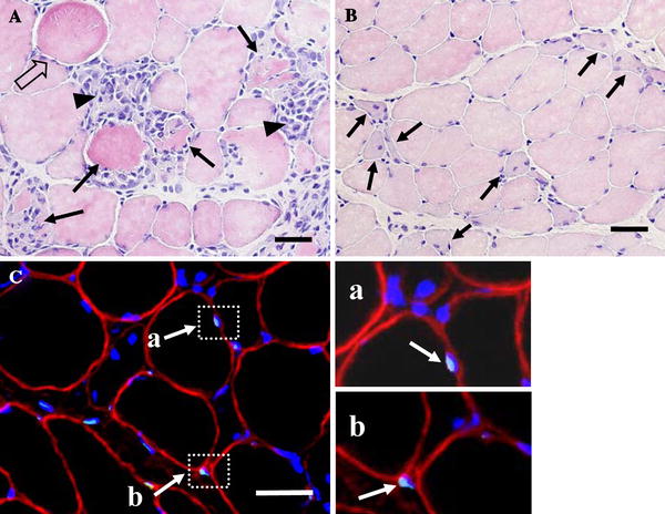
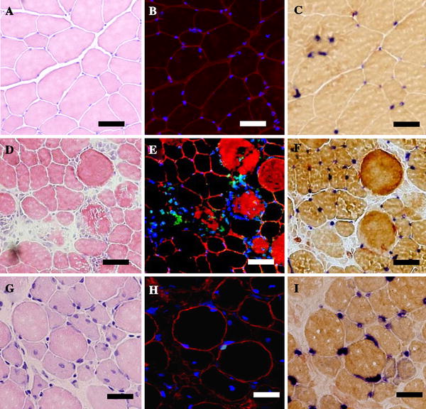
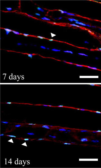
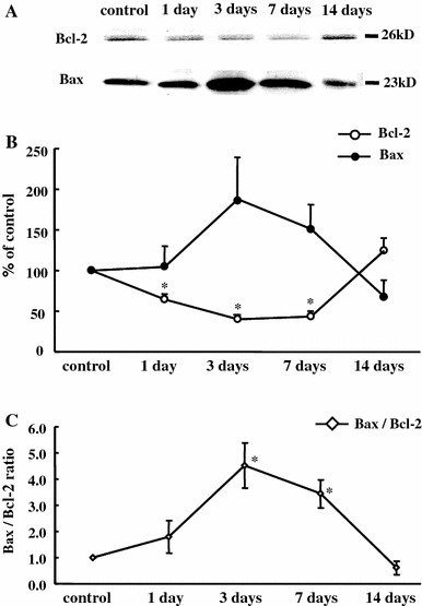
Eccentric contractions (ECC) induce myofibrillar collapse, edema, and inflammation in muscle cells. Although apoptosis of myonuclei following ECC is activated during the inflammatory phase, the apoptosis response of the regenerative phase remains to be elucidated. The aim of the present study was to determine the inflammatory and regenerative phase of the apoptosis responses induced by ECC. In anesthetized rats, the tibialis anterior muscles were subjected to ECC repeated 40 times, evoked by surface electric stimulation (100 Hz, 10 V) with mechanical muscle stretch. Apoptosis was examined in the control group and in groups 1, 3, 7, and 14 days after ECC (each group, n = 4-6). Terminal deoxynucleotidyl transferase-mediated dUTP nick end-labeling (TUNEL)-positive myonuclei were assessed by further labeling with dystrophin staining and DAPI. The expression of proteins related to apoptosis (Bcl-2 and Bax) was examined by Western blot assay. At 1 and 3 days, focal edema and necrotic myofibers invaded by mononuclear phagocytes were present, whereas regenerated myofibers with central nuclei were detected at 7 and 14 days. The occurrence of TUNEL-positive myonuclei increased significantly at 7 (7.0 +/- 1.5%) and 14 days (5.6 +/- 0.6%) compared with control (0.9 +/- 0.5%). Further we found that myonuclear apoptosis was restricted to the subsarcolemmal space at 7 and 14 days and markedly absent from the central nucleus. The Bax/Bcl-2 ratio was significantly higher at 3 (4.5 +/- 0.9) and 7 days (3.4 +/- 0.5) after ECC. In conclusion, myofiber apoptotic responses following ECC are present not only in the inflammatory phase but also persist during the regenerative phase.
Figures

Similar articles
-
Role of nitric oxide in muscle regeneration following eccentric muscle contractions in rat skeletal muscle.J Physiol Sci. 2013 Jul;63(4):263-70. doi: 10.1007/s12576-013-0262-y. Epub 2013 Apr 21. J Physiol Sci. 2013. PMID: 23606218 Free PMC article.
-
Loss of dystrophin and some dystrophin-associated proteins with concomitant signs of apoptosis in rat leg muscle overworked in extension.Acta Neuropathol. 2000 Dec;100(6):618-26. doi: 10.1007/s004010000231. Acta Neuropathol. 2000. PMID: 11078213
-
Cell death in denervated skeletal muscle is distinct from classical apoptosis.Anat Rec. 2000 Mar 1;258(3):305-18. doi: 10.1002/(SICI)1097-0185(20000301)258:3<305::AID-AR10>3.0.CO;2-A. Anat Rec. 2000. PMID: 10705351
-
Naturally occurring cell death during postnatal development of rat skeletal muscle.Muscle Nerve. 2002 Dec;26(6):777-83. doi: 10.1002/mus.10268. Muscle Nerve. 2002. PMID: 12451601
-
Apoptosis of myofibres and satellite cells: exercise-induced damage in skeletal muscle of the mouse.Neuropathol Appl Neurobiol. 1998 Dec;24(6):518-31. doi: 10.1046/j.1365-2990.1998.00149.x. Neuropathol Appl Neurobiol. 1998. PMID: 9888162
Cited by
-
Alterations of biochemical marker levels and myonuclear numbers in rat skeletal muscle after ischemia-reperfusion.Mol Cell Biochem. 2013 Jan;373(1-2):11-8. doi: 10.1007/s11010-012-1470-0. Epub 2012 Oct 13. Mol Cell Biochem. 2013. PMID: 23065010
-
Attenuated Oxidative Stress following Acute Exhaustive Swimming Exercise Was Accompanied with Modified Gene Expression Profiles of Apoptosis in the Skeletal Muscle of Mice.Oxid Med Cell Longev. 2016;2016:8381242. doi: 10.1155/2016/8381242. Epub 2016 Apr 10. Oxid Med Cell Longev. 2016. PMID: 27143996 Free PMC article.
-
Characteristics of nuclear architectural abnormalities of myotubes differentiated from LmnaH222P/H222P skeletal muscle cells.In Vitro Cell Dev Biol Anim. 2024 Aug;60(7):781-792. doi: 10.1007/s11626-024-00915-1. Epub 2024 May 9. In Vitro Cell Dev Biol Anim. 2024. PMID: 38724872
-
Serum-derived extracellular vesicles (EVs) impact on vascular remodeling and prevent muscle damage in acute hind limb ischemia.Sci Rep. 2017 Aug 15;7(1):8180. doi: 10.1038/s41598-017-08250-0. Sci Rep. 2017. PMID: 28811546 Free PMC article.
-
Role of nitric oxide in muscle regeneration following eccentric muscle contractions in rat skeletal muscle.J Physiol Sci. 2013 Jul;63(4):263-70. doi: 10.1007/s12576-013-0262-y. Epub 2013 Apr 21. J Physiol Sci. 2013. PMID: 23606218 Free PMC article.
References
-
- Allen DL, Linderman JK, Roy RR, Bigbee AJ, Grindeland RE, Mukku V, Edgerton VR. Apoptosis: a mechanism contributing to remodeling of skeletal muscle in response to hindlimb unweighting. Am J Physiol. 1997;273:C579–C587. - PubMed
MeSH terms
Substances
LinkOut - more resources
Full Text Sources
Research Materials
