

TLR8-mediated activation of human monocytes inhibits TL1A expression
- PMID: 19637197
- PMCID: PMC2839407
- DOI: 10.1002/eji.200939216
TLR8-mediated activation of human monocytes inhibits TL1A expression
Abstract
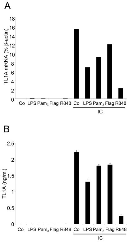
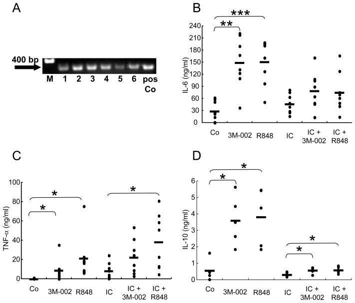
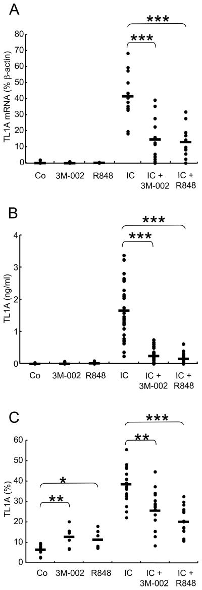
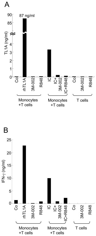
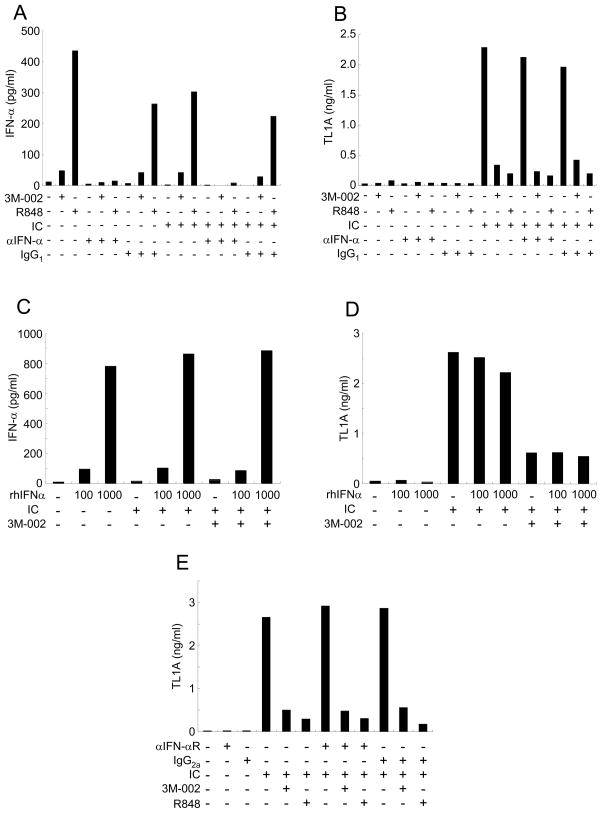
TLR play important roles in inflammation and innate immune response to pathogens. TLR8 recognizes ssRNA and induces NF-kappaB via MyD88 signaling. TL1A is a member of the TNF superfamily that markedly enhances IFN-gamma production by IL-12/IL-18-stimulated peripheral and mucosal CD4(+) T cells. TL1A expression is increased in the mucosa of patients with inflammatory bowel disease and is considered a key mediator of Crohn's disease (CD). We have previously shown that TL1A is strongly induced by immune complexes (IC) but not TLR ligands in antigen-presenting cells. However, a potential interaction between these pro-inflammatory signaling pathways has not been investigated. IC-induced TL1A expression of monocytes was potently inhibited by a TLR8 or TLR7/8 ligand (R848) in a dose-dependent manner. Furthermore, when co-cultured with CD4(+) T cells, TLR8 ligands inhibited TL1A production, resulting in almost complete inhibition of IFN-gamma production by the CD4(+) T cells. Furthermore, we demonstrate that IFN-alpha is not required for this suppressive effect by TLR8 signaling. Our data demonstrate for the first time a direct interaction between TLR and TL1A signaling pathways. TLR8 activation may be an important, novel pathway for targeted treatment of Th1-mediated diseases, such as CD.
Conflict of interest statement
The authors declare no competing financial interests.
Figures
Similar articles
-
The T cell costimulator TL1A is induced by FcgammaR signaling in human monocytes and dendritic cells.J Immunol. 2007 Apr 1;178(7):4033-8. doi: 10.4049/jimmunol.178.7.4033. J Immunol. 2007. PMID: 17371957
-
Microbial induction of inflammatory bowel disease associated gene TL1A (TNFSF15) in antigen presenting cells.Eur J Immunol. 2009 Nov;39(11):3239-50. doi: 10.1002/eji.200839087. Eur J Immunol. 2009. PMID: 19839006 Free PMC article.
-
Activation of Human γδ T Cells: Modulation by Toll-Like Receptor 8 Ligands and Role of Monocytes.Cells. 2020 Mar 13;9(3):713. doi: 10.3390/cells9030713. Cells. 2020. PMID: 32183240 Free PMC article.
-
Tumor Necrosis Factor-like Cytokine TL1A and Its Receptors DR3 and DcR3: Important New Factors in Mucosal Homeostasis and Inflammation.Inflamm Bowel Dis. 2015 Oct;21(10):2441-52. doi: 10.1097/MIB.0000000000000492. Inflamm Bowel Dis. 2015. PMID: 26099067 Review.
-
TL1A and DR3, a TNF family ligand-receptor pair that promotes lymphocyte costimulation, mucosal hyperplasia, and autoimmune inflammation.Immunol Rev. 2011 Nov;244(1):188-96. doi: 10.1111/j.1600-065X.2011.01068.x. Immunol Rev. 2011. PMID: 22017439 Free PMC article. Review.
Cited by
-
Structure-Based Design of Human TLR8-Specific Agonists with Augmented Potency and Adjuvanticity.J Med Chem. 2015 Oct 8;58(19):7833-49. doi: 10.1021/acs.jmedchem.5b01087. Epub 2015 Sep 22. J Med Chem. 2015. PMID: 26351878 Free PMC article.
-
Anti-TLR7 Antibody Protects Against Lupus Nephritis in NZBWF1 Mice by Targeting B Cells and Patrolling Monocytes.Front Immunol. 2021 Nov 11;12:777197. doi: 10.3389/fimmu.2021.777197. eCollection 2021. Front Immunol. 2021. PMID: 34868046 Free PMC article.
-
TL1A (TNFSF15) and DR3 (TNFRSF25): A Co-stimulatory System of Cytokines With Diverse Functions in Gut Mucosal Immunity.Front Immunol. 2019 Mar 27;10:583. doi: 10.3389/fimmu.2019.00583. eCollection 2019. Front Immunol. 2019. PMID: 30972074 Free PMC article. Review.
-
New insights into the dichotomous role of innate cytokines in gut homeostasis and inflammation.Cytokine. 2012 Sep;59(3):451-9. doi: 10.1016/j.cyto.2012.06.014. Epub 2012 Jul 12. Cytokine. 2012. PMID: 22795953 Free PMC article. Review.
-
Synergistic Activity of Second Mitochondrial-Derived Activator of Caspases Mimetic with Toll-like Receptor 8 Agonist Reverses HIV-1-Latency and Enhances Antiviral Immunity.Int J Mol Sci. 2025 Mar 13;26(6):2575. doi: 10.3390/ijms26062575. Int J Mol Sci. 2025. PMID: 40141220 Free PMC article.
References
-
- Migone TS, Zhang J, Luo X, Zhuang L, Chen C, Hu B, Hong JS, Perry JW, Chen SF, Zhou JX, Cho YH, Ullrich S, Kanakaraj P, Carrell J, Boyd E, Olsen HS, Hu G, Pukac L, Liu D, Ni J, Kim S, Gentz R, Feng P, Moore PA, Ruben SM, Wei P. TL1A is a TNF-like ligand for DR3 and TR6/DcR3 and functions as a T cell costimulator. Immunity. 2002;16:479–492. - PubMed
-
- Papadakis KA, Prehn JL, Landers C, Han Q, Luo X, Cha SC, Wei P, Targan SR. TL1A synergizes with IL-12 and IL-18 to enhance IFN-gamma production in human T cells and NK cells. J Immunol. 2004;172:7002–7007. - PubMed
-
- Prehn JL, Mehdizadeh S, Landers CJ, Luo X, Cha SC, Wei P, Targan SR. Potential role for TL1A, the new TNF-family member and potent costimulator of IFN-gamma, in mucosal inflammation. Clin Immunol. 2004;112:66–77. - PubMed
-
- Papadakis KA, Zhu D, Prehn JL, Landers C, Avanesyan A, Lafkas G, Targan SR. Dominant role for TL1A/DR3 pathway in IL-12 plus IL-18-induced IFN-gamma production by peripheral blood and mucosal CCR9+ T lymphocytes. J Immunol. 2005;174:4985–4990. - PubMed
-
- Bamias G, Martin C, 3rd, Marini M, Hoang S, Mishina M, Ross WG, Sachedina MA, Friel CM, Mize J, Bickston SJ, Pizarro TT, Wei P, Cominelli F. Expression, localization, and functional activity of TL1A, a novel Th1-polarizing cytokine in inflammatory bowel disease. J Immunol. 2003;171:4868–4874. - PubMed
Publication types
MeSH terms
Substances
Grants and funding
LinkOut - more resources
Full Text Sources
Other Literature Sources
Research Materials
Miscellaneous
