

Absence of beta3 integrin accelerates early skeletal repair
- PMID: 19637214
- PMCID: PMC2811376
- DOI: 10.1002/jor.20955
Absence of beta3 integrin accelerates early skeletal repair
Abstract
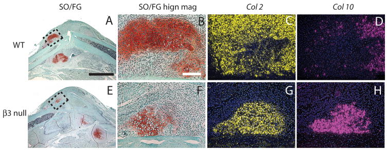
Integrins are heterodimeric transmembrane proteins that mediate cell-matrix interactions and modulate cell behavior. Beta3 subunit is a component of alphaIIbeta3 and alphaVbeta3 integrins. In this study, we first determined that beta3 transcripts are expressed by cells within fracture calluses at 7 and 10 days after injury in a mouse model. We then analyzed fracture healing in mice deficient of beta3 integrin with molecular, histomorphometric, and biomechanical techniques. We found that lack of beta3 integrin results in an extended bleeding time and leads to more bone formation and accelerated cartilage maturation at 7 days after injury. However, beta3 deficiency does not appear to affect later fracture healing. At days 14 and 21, histological appearance or biomechanical properties of fracture calluses are similar between wild type and mutant mice. We also found that altered fracture healing in beta3-null mice is not associated with accelerated angiogenesis, because no significant difference of length density and surface density of blood vessels in fracture limbs was detected at 3 days after injury between wild type and beta3-null mice. In conclusion, our findings demonstrate that beta3 integrin plays an important role during early fracture healing. Further research is required to determine the underlying mechanisms.
Figures


Similar articles
-
Delayed fracture healing in growth differentiation factor 5-deficient mice: a pilot study.Clin Orthop Relat Res. 2011 Oct;469(10):2915-24. doi: 10.1007/s11999-011-1912-0. Epub 2011 May 18. Clin Orthop Relat Res. 2011. PMID: 21590487 Free PMC article.
-
Diminished callus size and cartilage synthesis in alpha 1 beta 1 integrin-deficient mice during bone fracture healing.Am J Pathol. 2002 May;160(5):1779-85. doi: 10.1016/s0002-9440(10)61124-8. Am J Pathol. 2002. PMID: 12000729 Free PMC article.
-
Retarded chondrogenesis in transgenic mice with a type II collagen defect results in fracture healing abnormalities.Dev Dyn. 1994 Aug;200(4):340-9. doi: 10.1002/aja.1002000409. Dev Dyn. 1994. PMID: 7994081
-
CYR61 (CCN1) protein expression during fracture healing in an ovine tibial model and its relation to the mechanical fixation stability.J Orthop Res. 2006 Feb;24(2):254-62. doi: 10.1002/jor.20035. J Orthop Res. 2006. PMID: 16435358
-
Effect of age on vascularization during fracture repair.J Orthop Res. 2008 Oct;26(10):1384-9. doi: 10.1002/jor.20667. J Orthop Res. 2008. PMID: 18464248 Free PMC article.
Cited by
-
Bone regeneration materials and their application over 20 years: A bibliometric study and systematic review.Front Bioeng Biotechnol. 2022 Oct 5;10:921092. doi: 10.3389/fbioe.2022.921092. eCollection 2022. Front Bioeng Biotechnol. 2022. PMID: 36277397 Free PMC article.
-
Gene expression dynamics during bone healing and osseointegration.J Periodontol. 2011 Jul;82(7):1007-17. doi: 10.1902/jop.2010.100577. Epub 2010 Dec 13. J Periodontol. 2011. PMID: 21142982 Free PMC article.
-
Extracellular matrix-mimetic adhesive biomaterials for bone repair.J Biomed Mater Res A. 2011 Jan;96(1):261-72. doi: 10.1002/jbm.a.32979. Epub 2010 Nov 10. J Biomed Mater Res A. 2011. PMID: 21105174 Free PMC article. Review.
-
Effect of radiation on the expression of osteoclast marker genes in RAW264.7 cells.Mol Med Rep. 2012 Apr;5(4):955-8. doi: 10.3892/mmr.2012.765. Epub 2012 Jan 25. Mol Med Rep. 2012. PMID: 22294242 Free PMC article.
-
Osteocyte β3 integrin promotes bone mass accrual and force-induced bone formation in mice.J Orthop Translat. 2023 Jun 7;40:58-71. doi: 10.1016/j.jot.2023.05.001. eCollection 2023 May. J Orthop Translat. 2023. PMID: 37457310 Free PMC article.
References
-
- Quinn MJ, Byzova TV, Qin J, et al. Integrin alphaIIbbeta3 and its antagonism. Arterioscler Thromb Vasc Biol. 2003;23:945–52. - PubMed
-
- Zhao H, Kitaura H, Sands MS, et al. Critical role of beta3 integrin in experimental postmenopausal osteoporosis. J Bone Miner Res. 2005;20:2116–23. - PubMed
-
- Brooks PC, Clark RA, Cheresh DA. Requirement of vascular integrin alpha v beta 3 for angiogenesis. Science. 1994;264:569–71. - PubMed
Publication types
MeSH terms
Substances
Grants and funding
LinkOut - more resources
Full Text Sources
Medical
