

Patterned rhodopsin expression in R7 photoreceptors of mosquito retina: Implications for species-specific behavior
- PMID: 19637310
- PMCID: PMC2845463
- DOI: 10.1002/cne.22114
Patterned rhodopsin expression in R7 photoreceptors of mosquito retina: Implications for species-specific behavior
Abstract
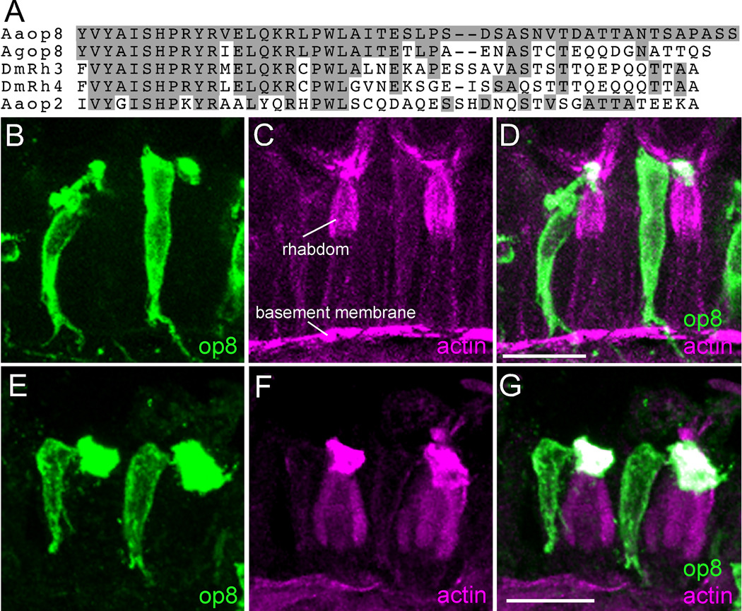
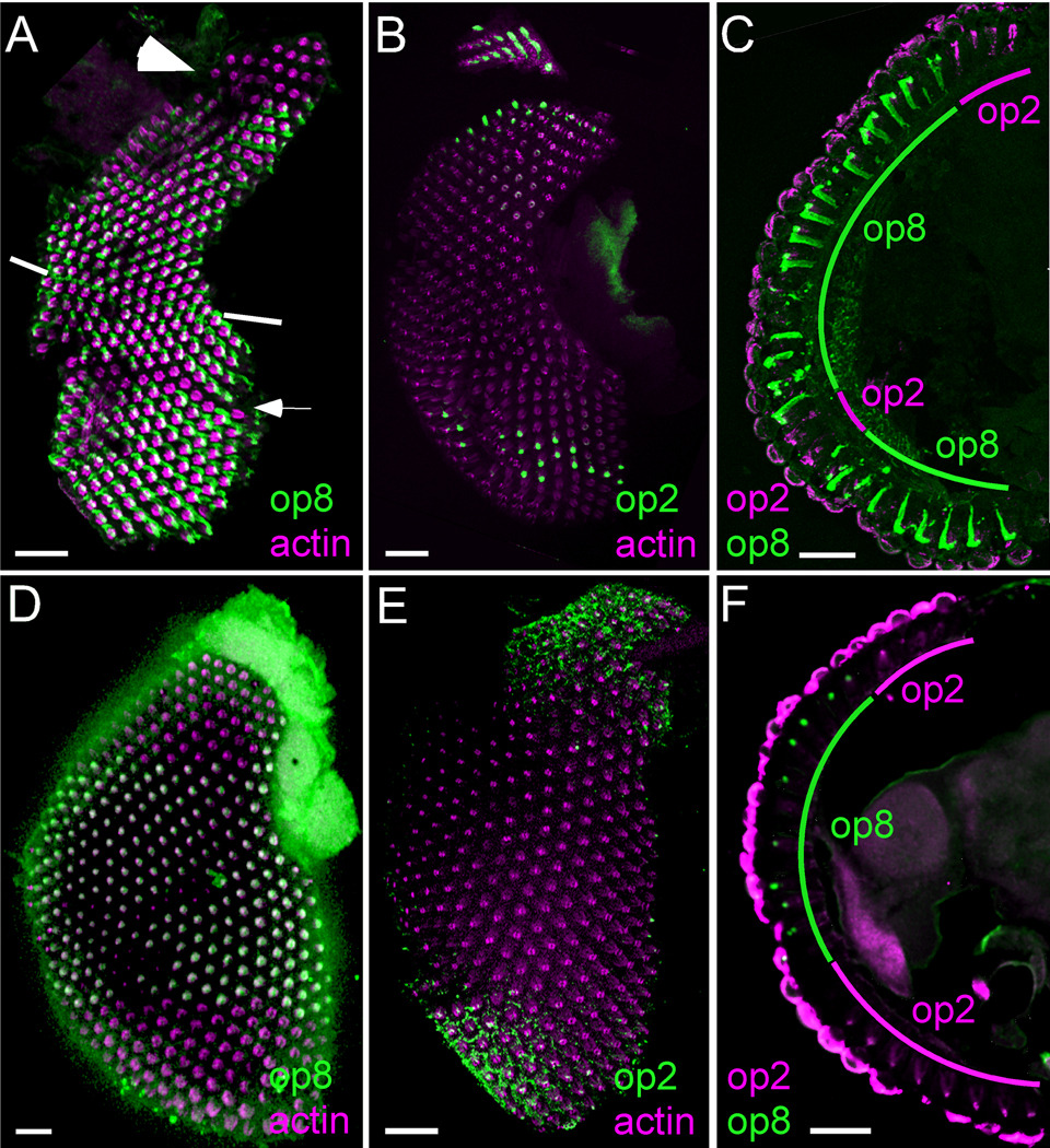
Visual perception of the environment plays an important role in many mosquito behaviors. Characterization of the cellular and molecular components of mosquito vision will provide a basis for understanding these behaviors. A unique feature of the R7 photoreceptors in Aedes aegypti and Anopheles gambiae is the extreme apical projection of their rhabdomeric membrane. We show here that the compound eye of both mosquitoes is divided into specific regions based on nonoverlapping expression of specific rhodopsins in these R7 cells. The R7 cells of the upper dorsal region of both mosquitoes express a long wavelength op2 rhodopsin family member. The lower dorsal hemisphere and upper ventral hemisphere of both mosquitoes express the UV-sensitive op8 rhodopsin. At the lower boundary of this second region, the R7 cells again express the op2 family rhodopsin. In Ae. aegypti, this third region is a horizontal stripe of one to three rows of ommatidia, and op8 is expressed in a fourth region in the lower ventral hemisphere. However, in An. gambiae the op2 family member expression is expanded throughout the lower region in the ventral hemisphere. The overall conserved ommatidial organization and R7 retinal patterning show these two species retain similar visual capabilities. However, the differences within the ventral domain may facilitate species-specific visual behaviors.
Figures


Similar articles
-
Rhodopsin coexpression in UV photoreceptors of Aedes aegypti and Anopheles gambiae mosquitoes.J Exp Biol. 2014 Mar 15;217(Pt 6):1003-8. doi: 10.1242/jeb.096347. Epub 2013 Dec 5. J Exp Biol. 2014. PMID: 24311804 Free PMC article.
-
Coexpression of spectrally distinct rhodopsins in Aedes aegypti R7 photoreceptors.PLoS One. 2011;6(8):e23121. doi: 10.1371/journal.pone.0023121. Epub 2011 Aug 8. PLoS One. 2011. PMID: 21858005 Free PMC article.
-
Expression and light-triggered movement of rhodopsins in the larval visual system of mosquitoes.J Exp Biol. 2015 May;218(Pt 9):1386-92. doi: 10.1242/jeb.111526. Epub 2015 Mar 6. J Exp Biol. 2015. PMID: 25750414 Free PMC article.
-
Molecular logic behind the three-way stochastic choices that expand butterfly colour vision.Nature. 2016 Jul 14;535(7611):280-4. doi: 10.1038/nature18616. Epub 2016 Jul 6. Nature. 2016. PMID: 27383790 Free PMC article.
-
Signaling mechanisms in induction of the R7 photoreceptor in the developing Drosophila retina.Bioessays. 1994 Apr;16(4):237-44. doi: 10.1002/bies.950160406. Bioessays. 1994. PMID: 8031300 Review.
Cited by
-
Expression of a homologue of a vertebrate non-visual opsin Opn3 in the insect photoreceptors.Philos Trans R Soc Lond B Biol Sci. 2022 Oct 24;377(1862):20210274. doi: 10.1098/rstb.2021.0274. Epub 2022 Sep 5. Philos Trans R Soc Lond B Biol Sci. 2022. PMID: 36058246 Free PMC article.
-
Deep conservation complemented by novelty and innovation in the insect eye ground plan.Proc Natl Acad Sci U S A. 2025 Jan 7;122(1):e2416562122. doi: 10.1073/pnas.2416562122. Epub 2024 Dec 30. Proc Natl Acad Sci U S A. 2025. PMID: 39793041 Free PMC article.
-
Spectral preferences of mosquitos are altered by odors.J Exp Biol. 2025 Jul 1;228(13):jeb250318. doi: 10.1242/jeb.250318. Epub 2025 Jul 9. J Exp Biol. 2025. PMID: 40539363 Free PMC article.
-
Rhodopsin management during the light-dark cycle of Anopheles gambiae mosquitoes.J Insect Physiol. 2014 Nov;70:88-93. doi: 10.1016/j.jinsphys.2014.09.006. Epub 2014 Sep 29. J Insect Physiol. 2014. PMID: 25260623 Free PMC article.
-
The sensory arsenal mosquitoes use to find us.Trends Parasitol. 2025 Jul;41(7):591-602. doi: 10.1016/j.pt.2025.05.004. Epub 2025 May 29. Trends Parasitol. 2025. PMID: 40447468 Free PMC article. Review.
References
-
- Ahmad ST, Natochin M, Artemyev NO, O'Tousa JE. The Drosophila rhodopsin cytoplasmic tail domain is required for maintenance of rhabdomere structure. Faseb J. 2007;21:449–455. - PubMed
-
- Brammer JD. The ultrastructure of the compound eye of a mosquito Aedes aegypti L. J Exp Zool. 1970;195:181–196.
-
- Briscoe AD, Chittka L. The evolution of color vision in insects. Annu Rev Entomol. 2001;46:471–510. - PubMed
-
- Clandinin TR, Zipursky SL. Making connections in the fly visual system. Neuron. 2002;35:827–841. - PubMed
-
- Day JF. Host-seeking strategies of mosquito disease vectors. J Am Mosq Control Assoc. 2005;21:17–22. - PubMed
Publication types
MeSH terms
Substances
Grants and funding
LinkOut - more resources
Full Text Sources
