. 2009 Oct 15;115(20):4655-66.
doi: 10.1002/cncr.24532.

Transport by SLC5A8 with subsequent inhibition of histone deacetylase 1 (HDAC1) and HDAC3 underlies the antitumor activity of 3-bromopyruvate

Affiliations

Transport by SLC5A8 with subsequent inhibition of histone deacetylase 1 (HDAC1) and HDAC3 underlies the antitumor activity of 3-bromopyruvate

Muthusamy Thangaraju et al. Cancer. .

Abstract

Background: 3-bromopyruvate is an alkylating agent with antitumor activity. It is currently believed that blockade of adenosine triphosphate production from glycolysis and mitochondria is the primary mechanism responsible for this antitumor effect. The current studies uncovered a new and novel mechanism for the antitumor activity of 3-bromopyruvate.

Methods: The transport of 3-bromopyruvate by sodium-coupled monocarboxylate transporter SMCT1 (SLC5A8), a tumor suppressor and a sodium (Na+)-coupled, electrogenic transporter for short-chain monocarboxylates, was studied using a mammalian cell expression and the Xenopus laevis oocyte expression systems. The effect of 3-bromopyruvate on histone deacetylases (HDACs) was monitored using the lysate of the human breast cancer cell line MCF7 and human recombinant HDAC isoforms as the enzyme sources. Cell viability was monitored by fluorescence-activated cell-sorting analysis and colony-formation assay. The acetylation status of histone H4 was evaluated by Western blot analysis.

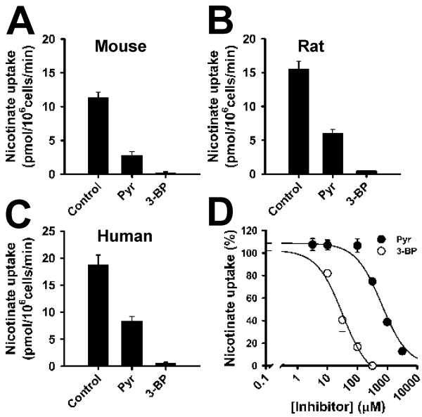
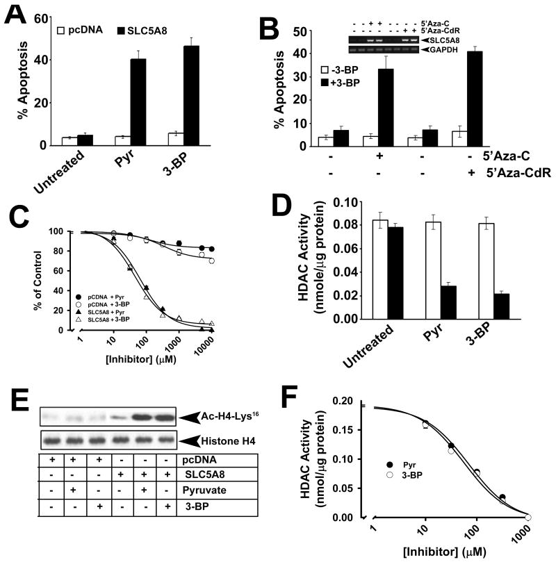
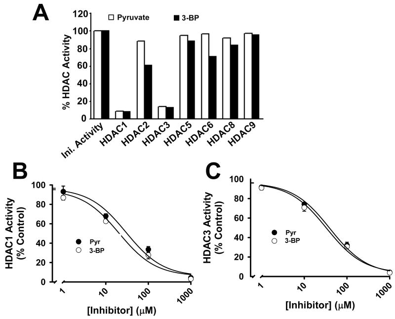
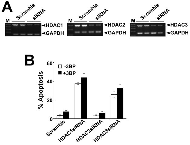
Results: 3-Bromopyruvate is a transportable substrate for SLC5A8, and that transport process is Na+-coupled and electrogenic. MCF7 cells did not express SLC5A8 and were not affected by 3-bromopyruvate. However, when transfected with SLC5A8 or treated with inhibitors of DNA methylation, these cells underwent apoptosis in the presence of 3-bromopyruvate. This cell death was associated with the inhibition of HDAC1/HDAC3. Studies with different isoforms of human recombinant HDACs identified HDAC1 and HDAC3 as the targets for 3-bromopyruvate.

Conclusions: 3-Bromopyruvate was transported into cells actively through the tumor suppressor SLC5A8, and the process was energized by an electrochemical Na+ gradient. Ectopic expression of the transporter in MCF7 cells led to apoptosis, and the mechanism involved the inhibition of HDAC1/HDAC3.


Figures

FIGURE 1
FIGURE 1
Interaction of 3-bromopyruvate with SLC5A8 in a mammalian cell expression system. Mouse (A), rat (B), and human (C) SLC5A8 cDNAs were expressed in HRPE cells, and the transport function was monitored by the uptake of nicotinate (15 μM) in the presence of Na+. Uptake values from vector-transfected cells were subtracted from uptake values from cDNA-transfected cells to determine SLC5A8-specific uptake. (A-C) SLC5A8-specific uptake of nicotinate in the absence (control) or presence of pyruvate (1 mM) or 3-bromopyruvate (1 mM). (D) Dose-response for the inhibition of SLC5A8-mediated nicotinate uptake by pyruvate and 3-bromopyruvate. Data represent means ± S. E. (n = 6-9). Pyr, pyruvate; 3-BP, 3-bromopyruvate.
FIGURE 2
FIGURE 2
Direct evidence for SLC5A8-mediated transport of 3-bromopyruvate. Human SLC5A8 was expressed in X. laevis oocytes and the transport function was monitored electrophysiologically using the two-microelectrode voltage-clamp technique. (A) Inward currents induced by pyruvate (1 mM) and 3-bromopyruvate (1 mM) in the presence (NaCl) or absence (NMDG chloride) of Na+. (B) Data from four different oocytes. In each oocyte, the current induced by pyruvate was taken as 100%. The value for pyruvate-induced currents in these four oocytes was 215 ± 45 nA.
FIGURE 3
FIGURE 3
Characteristics of SLC5A8-mediated transport of 3-bromopyruvate. (A) Relationship between substrate-induced currents and membrane potential. Concentration of pyruvate and 3-bromopyruvate was 1 mM. (B) Saturation kinetics for pyruvate-induced currents. (C) Saturation kinetics for 3-bromopyruvate-induced currents. (D) Na+-activation kinetics for 3-bromopyruvate-induced currents. Inset: Hill plot.
FIGURE 4
FIGURE 4
Apoptosis induced in MCF7 cells by 3-bromopyruvate is dependent on SLC5A8 and is associated with inhibition of HDACs. (A) MCF7 cells were transfected with either vector alone or human SLC5A8 cDNA, and then cultured in the absence or presence of pyruvate (1 mM) or 3-bromopyruvate (1 mM) for 48 h. Apoptosis was monitored by FACS. (B) MCF7 cells were treated with 5′-azacytidine (5′-Aza-C) or 5′-azadeoxycytidine (5′-Aza-CdR) for 72 h and then cultured in the absence or presence of 3-bromopyruvate as described in (A) and apoptosis was monitored by FACS. RNA prepared from these cells was used for RT-PCR to monitor SLC5A8 expression. (C) Dose-response for growth arrest and/or cell death induced by pyruvate and 3-bromopyruvate in vector-transfected and SLC5A8 cDNA-transfected MCF7 cells. Cell viability was monitored by colony formation assay. (D) Cells were treated as described for (A) and the cell lysates were used as the source of HDAC activity. (E) Cells were treated as described for (A) and the cell lysates were used for Western blot to determine the acetylation status of histone H4-Lys. (F) Dose-response for the inhibition of HDACs from MCF7 cells by pyruvate and 3-bromopyruvate. Data are means ± S. E. (n = 4).
FIGURE 5
FIGURE 5
Inhibition of human recombinant HDAC isoforms by pyruvate and 3-bromopyruvate. (A) Activity of individual HDAC isoforms was measured in the absence or presence of pyruvate (1 mM) or 3-bromopyruvate (1 mM). (B, C) Dose-response for inhibition of HDAC1 and HDAC3 by pyruvate and 3-bromopyruvate. Data are means ± S. E. (n = 3).
FIGURE 6
FIGURE 6
Relevance of inhibition of HDAC1 and HDAC3 to 3-bromopyruvate-induced apoptosis in MCF7 cells. MCF7 cells were transfected with HDAC1-, HDAC2- and HDAC3-specific siRNAs, and then incubated with or without 3-bromopyruvate (1 mM) for 48 h. A scrambled siRNA was used as a negative control. (A) RNA was extracted from these cells, and expression of HDAC1, HDAC2 and HDAC3 was monitored by RT-PCR using isoform-specific primers. GAPDH was used as a loading control. (B) Cells were treated as described in (A) and then used for FACS analysis to monitor apoptosis. Data are means ± S. E. (n = 3).

References

    1. Stubbs M, McSheehy PMJ, Griffiths JR, Bashford CL. Causes and consequences of tumour acidity and implications for treatment. Mol Med Today. 2000;6:15–19. - PubMed
    1. Gatenby RA, Gillies RJ. Why do cancers have high aerobic glycolysis? Nat Rev Cancer. 2004;4:891–899. - PubMed
    1. Ristow M. Oxidative metabolism in cancer growth. Curr Opin Clin Nutr Metab Care. 2006;9:339–345. - PubMed
    1. Kim J, Dang CV. Cancer's molecular sweet tooth and the Warburg effect. Cancer Res. 2006;66:8927–8930. - PubMed
    1. Pedersen PL. The cancer cell's “power plants” as promising therapeutic targets: An overview. J Bioenerg Biomembr. 2007;39:1–12. - PubMed

Publication types

MeSH terms