

Defect in CEACAM family member expression in Crohn's disease IECs is regulated by the transcription factor SOX9
- PMID: 19637360
- PMCID: PMC3005567
- DOI: 10.1002/ibd.21023
Defect in CEACAM family member expression in Crohn's disease IECs is regulated by the transcription factor SOX9
Abstract
Background: CEACAM1, CEACAM5, and CEACAM6 represent 3 of the CEACAM (carcinoembryonic antigen-related cell adhesion molecule) subfamily members expressed on intestinal epithelial cells (IECs). Deficiency in their expression, as seen in inflammatory bowel disease (IBD), results in the lack of activation of CD8+ regulatory T cells in the mucosa. Since CEACAM expression was shown to be regulated by the transcription factor SOX9, we sought to determine whether the defect in CEACAM expression in IBD was related to aberrant SOX9 expression.
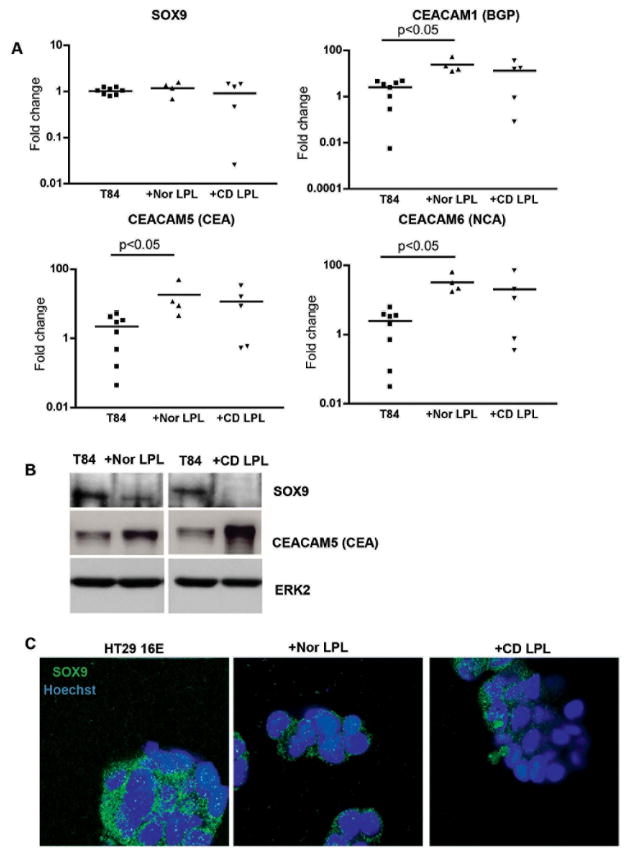
Methods: IECs and lamina propria lymphocytes (LPLs) were freshly isolated from colonic tissues. T84 and HT29 16E cells were cocultured with LPLs. SOX9 and CEACAM subfamily member expression was assessed by real-time polymerase chain reaction (PCR), Western blot, immunohistochemistry, and immunofluorescence.
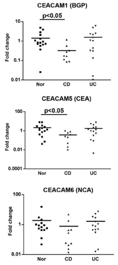
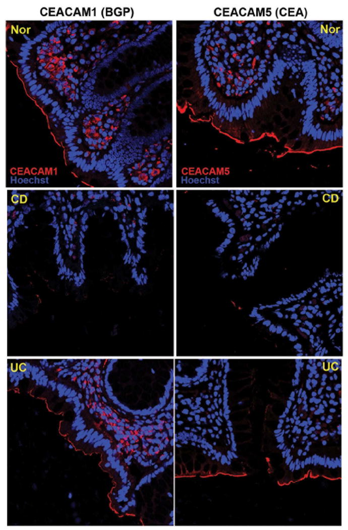
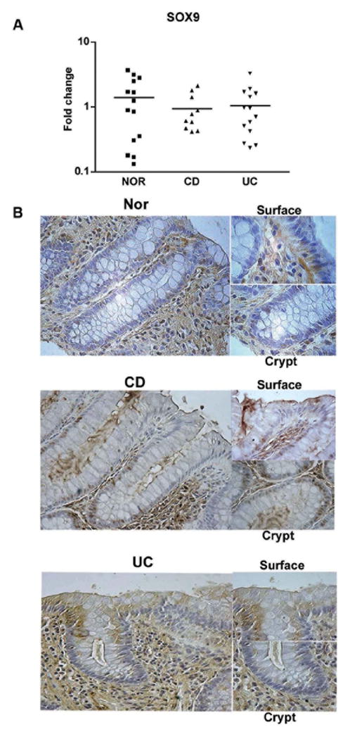
Results: In Crohn's disease (CD) but not in ulcerative colitis (UC), a significant reduction in mRNA and protein expression for CEACAM1 and 5 was noted; in contrast, no difference in SOX9 mRNA expression was seen. However, nuclear SOX9 immunostaining was increased in CD IECs. Furthermore, SOX9 protein was reduced in the cytoplasm of LPL-stimulated T84 and HT29 16E cells, while CEACAM5 expression was increased.
Conclusions: The defect in CEACAM family members in CD IECs appears to be related to the aberrant nuclear localization of SOX9. Changes in SOX9 expression in the CD mucosa relate to the local microenvironment and altered IEC:LPL crosstalk.
Copyright © 2009 Crohn's & Colitis Foundation of America, Inc.
Figures
References
-
- Allez M, Brimnes J, Dotan I, et al. Expansion of CD8+ T cells with regulatory function after interaction with intestinal epithelial cells. Gastroenterology. 2002;123:1516–1526. - PubMed
-
- Allez M, Brimnes J, Shao L, et al. Activation of a unique population of CD8(+) T cells by intestinal epithelial cells. Ann N Y Acad Sci. 2004;1029:22–35. - PubMed
-
- Allez M, Mayer L. Regulatory T cells: peace keepers in the gut. Inflamm Bowel Dis. 2004;10:666–676. - PubMed
Publication types
MeSH terms
Substances
Grants and funding
LinkOut - more resources
Full Text Sources
Medical
Research Materials
Miscellaneous