. 2009 Sep-Oct;6(5):1506-17.
doi: 10.1021/mp900081y.

Galactosylated LDL nanoparticles: a novel targeting delivery system to deliver antigen to macrophages and enhance antigen specific T cell responses

Affiliations

Galactosylated LDL nanoparticles: a novel targeting delivery system to deliver antigen to macrophages and enhance antigen specific T cell responses

Fang Wu et al. Mol Pharm. 2009 Sep-Oct.

Abstract

We aim to define the role of Kupffer cells in intrahepatic antigen presentation, using the selective delivery of antigen to Kupffer cells rather than other populations of liver antigen-presenting cells. To achieve this we developed a novel antigen delivery system that can target antigens to macrophages, based on a galactosylated low-density lipoprotein nanoscale platform. Antigen was delivered via the galactose particle receptor (GPr), internalized, degraded and presented to T cells. The conjugation of fluoresceinated ovalbumin (FLUO-OVA) and lactobionic acid with LDL resulted in a substantially increased uptake of FLUO-OVA by murine macrophage-like ANA1 cells in preference to NIH3T3 cells, and by primary peritoneal macrophages in preference to primary hepatic stellate cells. Such preferential uptake led to enhanced proliferation of OVA specific T cells, showing that the galactosylated LDL nanoscale platform is a successful antigen carrier, targeting antigen to macrophages but not to all categories of antigen presenting cells. This system will allow targeted delivery of antigen to macrophages in the liver and elsewhere, addressing the question of the role of Kupffer cells in liver immunology. It may also be an effective way of delivering drugs or vaccines directly at macrophages.


Figures

Figure 1
Figure 1
Mechanism of uptake of FLUO-OVA-GAL-LDL nanoparticles. The scheme represents localization of FLUO-OVA-GAL-LDL in endosomes/lysosomes after internalization through the GPr (galactose particle receptor) endocytosis pathway. Then the antigen OVA is degraded and transferred to the cytosol, processed to form a complex with MHC class I molecules, and subsequently presented to OVA specific OT-1 T cells, resulting in their proliferation.
Figure 2
Figure 2
Scheme of the conjugation method for the preparation of FLUO-OVA-GAL-LDL nanoparticles.
Figure 3
Figure 3
TEM images of (A) native LDL (B) FLUO-OVA-GAL-LDL nanoparticles, scale bar=100 nm.
Figure 4
Figure 4
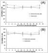
Cytotoxicity of FLUO-OVA-GAL-LDL towards ANA1 cells (A) and NIH3T3 cells (B). The ANA1 cells or NIH3T3 cells were treated with FLUO-OVA-GAL-LDL nanoparticles at different OVA concentration, or with free FLUO-OVA. Relative cell viability was expressed as a percentage of that in control cells treated with media alone. Comparable to free FLUO-OVA, FLUO-OVA-GAL-LDL showed minimal cytotoxicity towards either cell line after 24 h of incubation treatment.
Figure 5
Figure 5
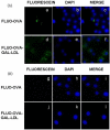
Specific delivery of FLUO-OVA-GAL-LDL to ANA1 cells. Representative fluorescent micrographs of FLUO-OVA visualized by fluorescein (green, a, d, g, j) demonstrate FLUO-OVA uptake by ANA1 cells (A) and lack of such uptake by NIH3T3 cells (B) after 5 h of incubation. The DNA in nuclei was stained with DAPI (blue, b, e, h, k). Micrographs c, f, i and l are the merge images of the fluorescein and DAPI images on the same row.
Figure 6
Figure 6
(A) Mean fluorescence intensity measured by flow cytometry to indicate the uptake of FLUO-OVA by ANA1 cells or NIH3T3 cells after 5 h incubation with 2 μg/mL of free FLUO-OVA or with FLUO-OVA-GAL-LDL, made using different galactosylation ratios. (B) The uptake of FLUO-OVA-GAL-LDL was inhibited by a 10-fold excess of GAL-LDL relative to a PBS control, but not by a 10-fold excess of LDL. The MFI of fluorescence was compared in ANA1 cells treated with FLUO-OVA-GAL-LDL after pretreatment with either PBS, or GAL-LDL, or LDL alone. The significances of the differences were evaluated using ANOVA test. Results are expressed as average values ±S.D. from three independent experiments. Significant differences with P<0.05 are indicated *, while N.S. indicates no significant difference. (C) Mean fluorescence intensity measured by flow cytometry to indicate the uptake of FLUO-OVA by primary peritoneal macrophages or hepatic stellate cells after 5 h incubation with 2 μg/mL of free FLUO-OVA, versus FLUO-OVA-GAL-LDL prepared using different galactosylation ratios.
Figure 7
Figure 7
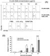
(A) Representative histogram of CFSE-labeled OT-1 T cells after 5 days of co-culture with peritoneal macrophages pulsed for 14h with different dose of FLUO-OVA or FLUO-OVA-GAL-LDL nanoparticles. Histograms are gated on Vα2+CD8+ cells. Proliferation was detected as dilution of CFSE on OT-1 cells. Control wells were pulsed with PBS, LDL or SINFEKL before OT-1 cell addition. (B) Percentage of divided OT-1 T cell obtained after 5 days of co-culture with peritoneal macrophages pulsed for 14h with different dose of FLUO-OVA or FLUO-OVA-GAL-LDL nanoparticles. Results are expressed as average values ±S.D. from three independent experiments. Significance of differences in the percentage of divided T cells, in response to macrophages treated with FLUO-OVA versus FLUO-OVA-GAL-LDL was determined using ANOVA test: *, p<0.05, **, p<0.01, ***, p<0.001.

Similar articles

Cited by

References

    1. Mackay IR. Hepatoimmunology: a perspective. Immunol. Cell Biol. 2002;80(1):36–44. - PubMed
    1. Racanelli V, Rehermann B. The liver as an immunological organ. Hepatology. 2006;43(2 Suppl 1):S54–62. - PubMed
    1. Bertolino P, McCaughan GW, Bowen DG. Role of primary intrahepatic T-cell activation in the ‘liver tolerance effect’. Immunol. Cell Biol. 2002;80(1):84–92. - PubMed
    1. Dong L, Zuo L, Xia S, Gao S, Zhang C, Chen J, Zhang J. Reduction of liver tumor necrosis factor-alpha expression by targeting delivery of antisense oligonucleotides into Kupffer cells protects rats from fulminant hepatitis. J. Gene Med. 2009;11(3):229–239. - PubMed
    1. van Rooijen N. Liposomes for targeting of antigens and drugs: immunoadjuvant activity and liposome-mediated depletion of macrophages. J. Drug Target. 2008;16(7):529–534. - PubMed

Publication types

MeSH terms