. 2009 Aug 19;131(32):11458-70.
doi: 10.1021/ja902140m.

C-lysine conjugates: pH-controlled light-activated reagents for efficient double-stranded DNA cleavage with implications for cancer therapy

Affiliations

C-lysine conjugates: pH-controlled light-activated reagents for efficient double-stranded DNA cleavage with implications for cancer therapy

Wang-Yong Yang et al. J Am Chem Soc. .

Abstract

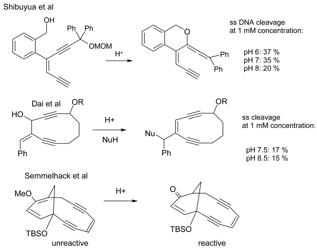
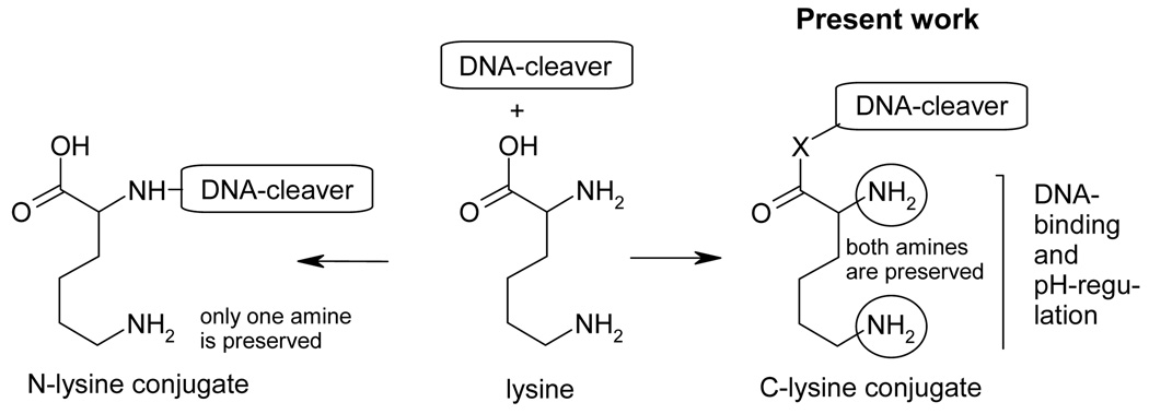
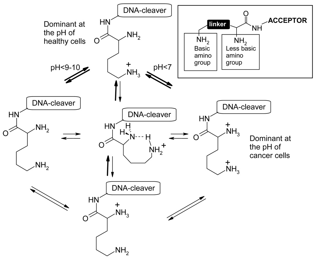
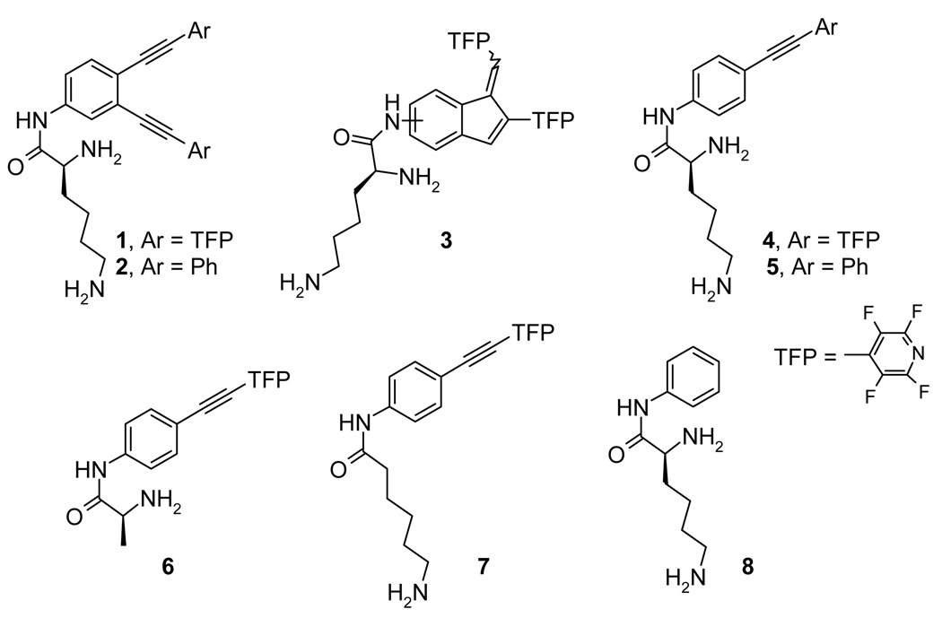
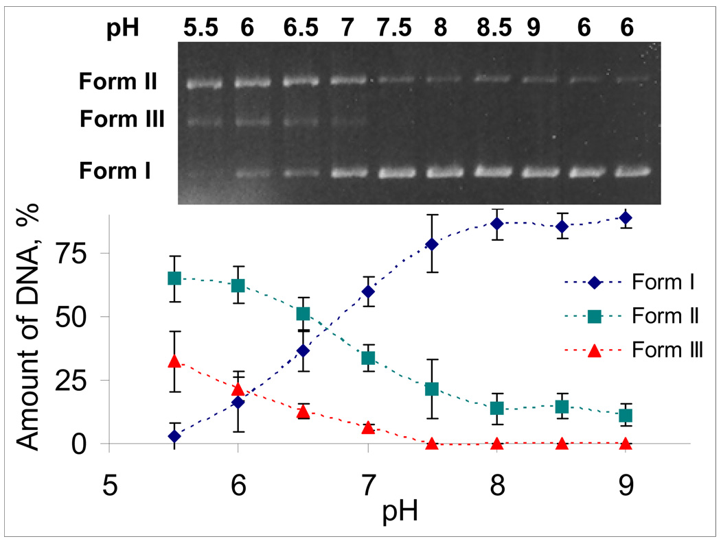
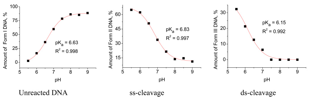
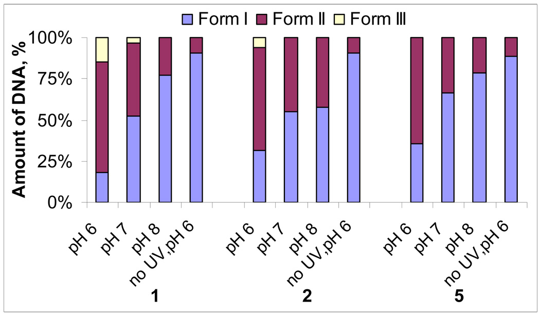
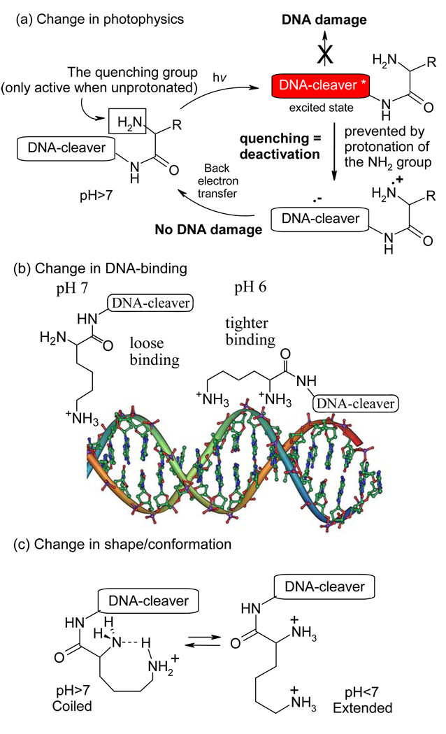
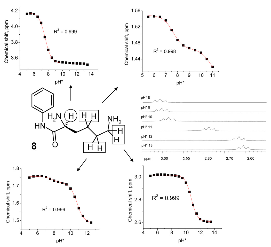
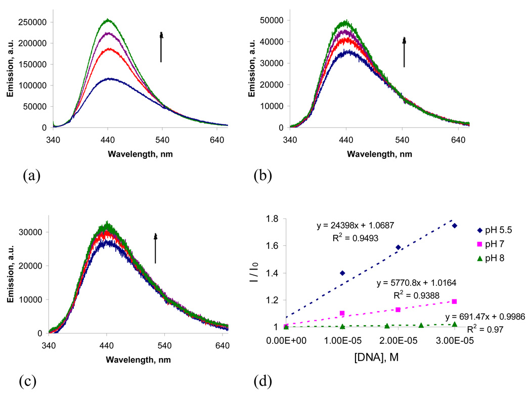
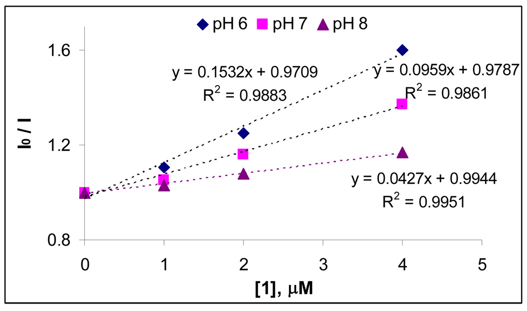
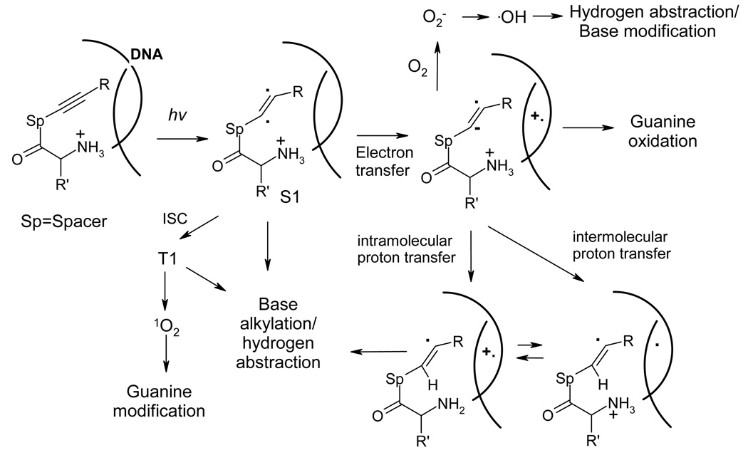
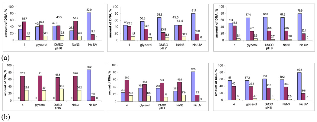
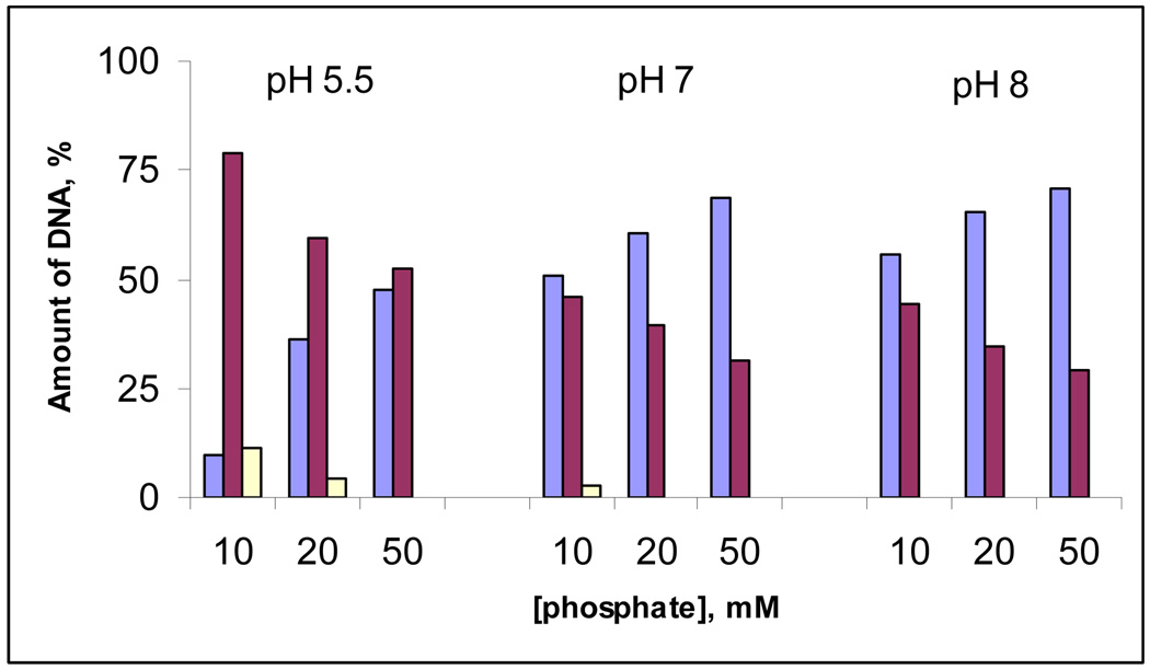
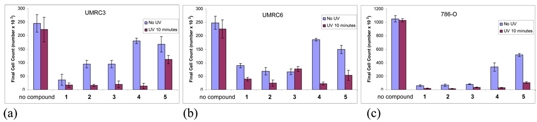
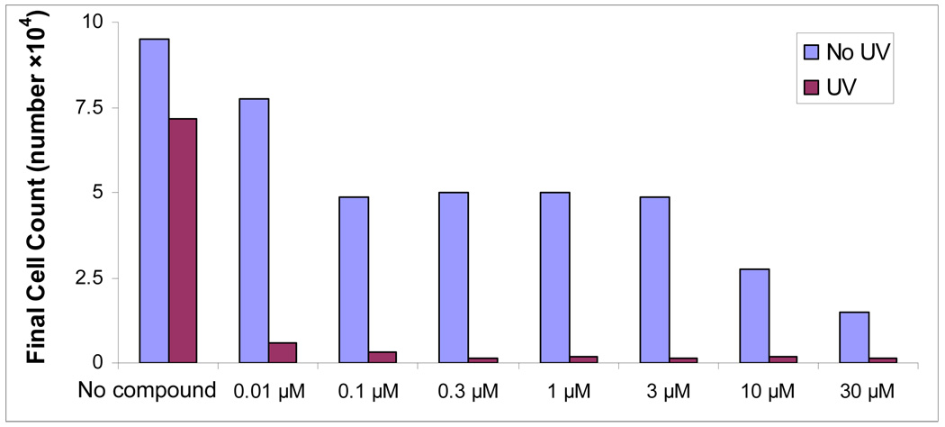
Double-stranded DNA cleavage of light-activated lysine conjugates is strongly enhanced at the slightly acidic pH (<7) suitable for selective targeting of cancer cells. This enhancement stems from the presence of two amino groups of different basicities. The first amino group plays an auxiliary role by enhancing solubility and affinity to DNA, whereas the second amino group, which is positioned next to the light-activated DNA cleaver, undergoes protonation at the desired pH threshold. This protonation results in two synergetic effects which account for the increased DNA-cleaving ability at the lower pH. First, lysine conjugates show tighter binding to DNA at the lower pH, which is consistent with the anticipated higher degree of interaction between two positively charged ammonium groups with the negatively charged phosphate backbone of DNA. Second, the unproductive pathway which quenches the excited state of the photocleaver through intramolecular electron transfer is eliminated once the donor amino group next to the chromophore is protonated. Experiments in the presence of traps for diffusing radicals show that reactive oxygen species do not contribute significantly to the mechanism of DNA cleavage at the lower pH, which is indicative of tighter binding to DNA under these conditions. This feature is valuable not only because many solid tumors are hypoxic but also because cleavage which does not depend on diffusing species is more localized and efficient. Sequence-selectivity experiments suggest combination of PET and base alkylation as the chemical basis for the observed DNA damage. The utility of these molecules for phototherapy of cancer is confirmed by the drastic increase in toxicity of five conjugates against cancer cell lines upon photoactivation.


Figures

Figure 1
Figure 1
Approaches to pH-activated enediynes based on acid-catalyzed transformations of unreactive prodrugs.
Figure 2
Figure 2
Literature examples of pH-controlled amino enediynes.
Figure 3
Figure 3
Two types of lysine conjugates
Figure 4
Figure 4
Design of pH-dependent DNA-cleavers based on different stages of protonation of the lysine side chain. The dominant protonation pathway is indicated by the bold arrows.
Figure 5
Figure 5
Structures of lysine conjugates 1– 5 and related control compounds 6 – 8.
Figure 6
Figure 6
Plasmid relaxation assays with conjugate 4 (15 µM) and pBR 322 plasmid DNA (30 µM/bp) in 20 mM sodium phosphate buffer after 3 minutes of irradiation. Form I = intact supercoiled DNA, Form II = relaxed form (ss cleavage), Form III = linear form (ds cleavage). Lanes 1–8: from pH 5.5 to pH 9, lane 9: no compound + UV, lane 10: compound + no UV. Cleavage data were quantified through densitometry. Reported values represent the average of four experiments.
Figure 7
Figure 7
Percentage of unreacted DNA (Form I), single-stranded cleavage (Form II) and double-stranded cleavage (Form III) as a function of pH, and the apparent pKa values corresponding to these plots.
Figure 8
Figure 8
Quantified plasmid relaxation assays with 15 µM of compounds 1, 2, 5 and 30 µM/bp of pBR 322 plasmid DNA in 20 mM sodium phosphate buffer at pH 6, 7 and 8 after 3 min. irradiation.
Figure 9
Figure 9
Plasmid relaxation assays for DNA photocleavage with 15 µM of compounds 4, 6 and 7 and pBR322 plasmid DNA (30 µM/bp) in 20 mM sodium phosphate buffer (pH 6, 7, 8) after 10 min of UV irradiation. Quantified cleavage data are presented on the left, the original gels are on the right.
Figure 10
Figure 10
Quantified plots of pBR 322 plasmid (30 µM/bp) relaxation assays for 10 µM of compound 4 in 20 mM sodium phosphate buffer at pH 6 (a), 7 (b) and 8 (c) as a function of the irradiation time. Plots on the bottom show effect of UV on the DNA without the conjugate at different pH. Plots on the top illustrate reactivity in the presence of compound 4.
Figure 11
Figure 11
Three possible mechanisms for pH-regulated DNA modification by lysine conjugates.
Figure 12
Figure 12
1H-NMR spectra of α- and ε- hydrogens in compound 8 (5 mM) in D2O at pD 5, 6, 7, 8, 9, 10, 11, 12, 13, and chemical shift-pD titration data for α- and ε- hydrogens in compound 8. These pKa values change to 7.44 and 10.40 when correction for going from pKa H* to pKa is applied
Figure 13
Figure 13
Fluorescence spectra of compound 4 (10 µM) at pH 4.1, 5.0, 6.1, 6.5, 6.9, 7.2, 8.0, 9.0, and 9.5. Insert: Quantified changes in fluorescence intensity as a function of pH and their fit to the Henderson-Hasselbalch equation for an acid-base equilibrium involving one amino group.
Figure 14
Figure 14
The pH-dependent lifetimes of the singlet excited state for conjugates 4 and 6 (10 µM), measured in 20 mM sodium phosphate buffer.
Figure 15
Figure 15
Proton affinities (in kcal/mol) of α- and ε-nitrogen lone pairs of diamines 4 and 8 at the B3LYP/6-31G(d, p) level of theory with PCM solvation in H2O.
Figure 16
Figure 16
UV-Vis absorbance titrations of 4 (10 µM) in 20 mM sodium phosphate buffer with 0, 1, 2, 3 equivalents of 30 μM/bp CT DNA at pH 5.5 (a), pH 7 (b) and pH 8 (c).
Figure 17
Figure 17
Emission titrations of 4 (10µM) in 20 mM sodium phosphate buffer with 0, 10, 20, 30 μM of CT DNA at pH 5.5 (a), pH 7 (b) and pH 8 (c). The initial slopes of plots illustrating changes in fluorescence of 4 as a function of DNA concentration (d).
Figure 18
Figure 18
Changes in fluorescence of ethidium bromide (10 µM, the excitation wavelength = 535 nm) in CT DNA (10 µM) as a function of displacement by compound 1
Figure 19
Figure 19
Summary of possible mechanistic alternatives for the observed DNA cleavage by amino acid / acetylene conjugates.
Figure 20
Figure 20
Effect of hydroxyl radical/singlet oxygen scavengers (10 mM) upon the efficiency of DNA cleavage at pH 6, 7 and 8 by conjugates 1 (a) and 4 (b) after 5 min. irradiation. Color coding: light blue – Form I, dark purple – Form II, light yellow – Form III.
Figure 21
Figure 21
Quantified Cleavage data of pBR 322 plasmid (30 µM/bp) relaxation assays of compound 4 (10 µM) at different concentrations of sodium phosphate buffer at pH 5.5, 7 and 8. Color coding: light blue – Form I, dark purple – Form II, light yellow – Form III.
Figure 22
Figure 22
Cell proliferation assays using three different RCC cell lines: (a) UMRC3, (b) UMRC6 and (c) 786-O, and compounds 1–5. RCC cells were resuspended in media and treated with UV radiation (350 nm) for 10 minutes with and without indicated compounds. Control cells were exposed to the indicated compound 1–5 at final concentration of 10 µM. Cell numbers determined after 72 hours as described in the SI Section.
Figure 23
Figure 23
LNCap cell proliferation assays with varying concentration of compound 4. The cells were exposed to UV for 10 minutes. Cells were counted 48 hours after the exposure. Full experimental details are described in the SI Section.

References

    1. Schnarr NA, Kennan A. Org. Lett. 2005;7:395. - PubMed
    2. Schnarr NA, Kennan AJ. J. Am. Chem. Soc. 2003;125:6364. - PubMed
    1. Rooth M, Shaw AM. J.Phys. Chem. C. 2007;111:15363.
    1. Xu H, Stampp SP, Rudkevich DM. M. Org. Lett. 2003;5:4583. - PubMed
    1. Choi HS, Huh KM, Ooya T, Yui N. J. Am. Chem. Soc. 2003;125:6350. - PubMed
    1. Martinez-Diaz M-V, Spencer N, Stoddart JF. Angew. Chem., Int. Ed. Engl. 1997;36:1904.
    2. Saha S, Stoddart JF. Chem. Soc. Rev. 2007;36:77. - PubMed
    3. Ashton PR, Ballardini R, Balzani V, Baxter I, Credi A, Fyfe MCT, Gandolfi MT, Gomez-Lopez M, Martinez-Diaz M-V, Piersanti A, Spencer N, Stoddart JF, Venturi M, White AJP, Williams DJ. J. Am. Chem. Soc. 1998;120:11932.
    4. Elizarov AM, Chang T, Chiu H-S, Stoddart JF. J. Org. Chem. 2002;67:9175. - PubMed
    5. Silvi S, Arduini A, Pochini A, Secchi A, Tomasulo M, Raymo FM, Baroncini M, Credi A. J. Am. Chem. Soc. 2007;129:13378. - PubMed
    6. Leigh DA, Thomson AR. Org. Lett. 2006;8:5377. - PubMed

Publication types