

Early mitochondrial dysfunction after cortical contusion injury
- PMID: 19637966
- PMCID: PMC2850255
- DOI: 10.1089/neu.2008.0857
Early mitochondrial dysfunction after cortical contusion injury
Abstract
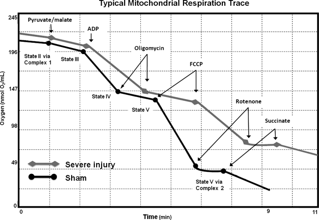
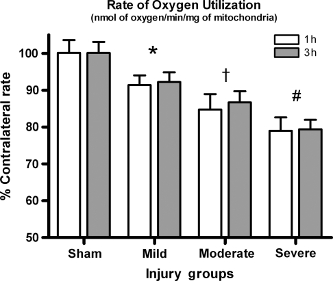
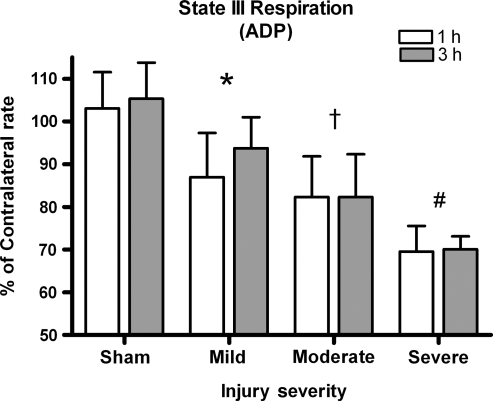
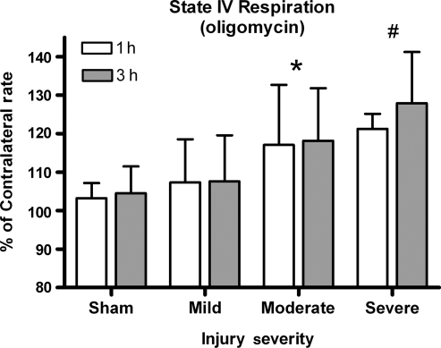
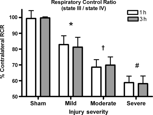
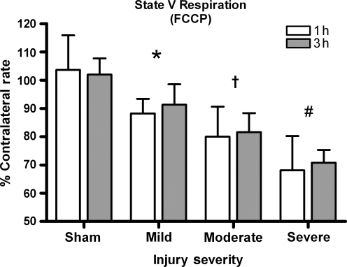
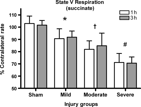
Following traumatic brain injury, mitochondria sustain structural and functional impairment, which contributes to secondary damage that can continue for days after the initial injury. The present study investigated mitochondrial bioenergetic changes in the rat neocortex at 1 and 3 h after mild, moderate, and severe injuries. Brains from young adult Sprague-Dawley rats were harvested from the injured and contralateral cortex to assess possible changes in mitochondrial respiration abilities following a unilateral cortical contusion injury. Differential centrifugation was used to isolate synaptic and extrasynaptic mitochondria from cortical tissue. Bioenergetics was assessed using a Clark-type electrode and results were graphed as a function of injury severity and time post-injury. Respiration was significantly affected by all injury severity levels compared to uninjured tissue. Complex 1- and complex 2-driven respirations were affected proportionally to the severity of the injury, indicating that damage to mitochondria may occur on a gradient. Total oxygen utilization, respiratory control ratio, ATP production, and maximal respiration capabilities were all significantly decreased in the injured cortex at both 1 and 3 h post-trauma. Although mitochondria displayed bioenergetic deficits at 1 h following injury, damage was not exacerbated by 3 h. This study stresses the importance of early therapeutic intervention and suggests a window of approximately 1-3 h before greater dysfunction occurs.
Figures
References
-
- Baldwin S.A. Scheff S.W. Intermediate filament change in astrocytes following mild cortical contusion. Glia. 1996;16:266–275. - PubMed
-
- Brustovetsky N. Brustovetsky T. Jemmerson R. Dubinsky J.M. Calcium-induced cytochrome c release from CNS mitochondria is associated with the permeability transition and rupture of the outer membrane. J. Neurochem. 2002;80:207–218. - PubMed
-
- Clark R.S. Nathaniel P.D. Zhang X. Dixon C.E. Alber S.M. Watkins S.C. Melick J.A. Kochanek P.M. Graham S.H. Boc-aspartyl(ome)-fluoromethylketone attenuates mitochondrial release of cytochrome C and delays brain tissue loss after traumatic brain injury in rats. J. Cereb. Blood Flow Metab. 2007;27:316–326. - PubMed
-
- Clausen T. Zauner A. Levasseur J.E. Rice A.C. Bullock R. Induced mitochondrial failure in the feline brain: Implications for understanding acute post-traumatic metabolic events. Brain Res. 2001;908:35–48. - PubMed
Publication types
MeSH terms
Grants and funding
LinkOut - more resources
Full Text Sources
