. 2009 Sep 25;284(39):26778-88.
doi: 10.1074/jbc.M109.032342. Epub 2009 Jul 28.

Insig-mediated, sterol-accelerated degradation of the membrane domain of hamster 3-hydroxy-3-methylglutaryl-coenzyme A reductase in insect cells

Affiliations

Insig-mediated, sterol-accelerated degradation of the membrane domain of hamster 3-hydroxy-3-methylglutaryl-coenzyme A reductase in insect cells

Andrew D Nguyen et al. J Biol Chem. .

Abstract

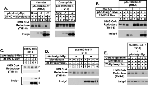
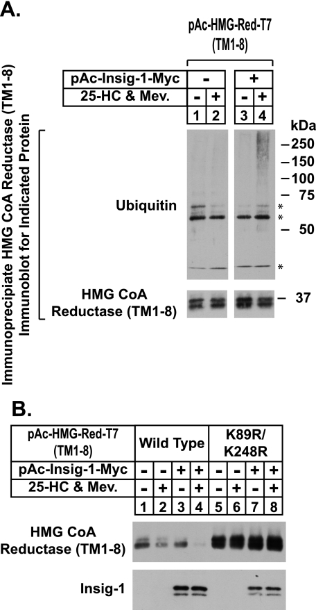
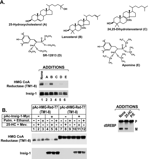
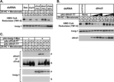
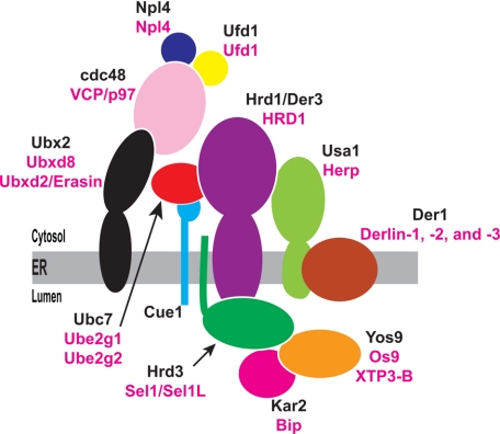
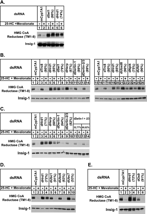
Sterol-accelerated degradation of the enzyme 3-hydroxy-3-methylglutaryl-coenzyme A reductase is one of several mechanisms through which cholesterol synthesis is controlled in mammalian cells. This degradation results from sterol-induced binding of the membrane domain of reductase to endoplasmic reticulum membrane proteins called Insig-1 and Insig-2, which are carriers of a ubiquitin ligase called gp78. The ensuing gp78-mediated ubiquitination of reductase is a prerequisite for its rapid, 26 S proteasome-mediated degradation from endoplasmic reticulum membranes, a reaction that slows a rate-limiting step in cholesterol synthesis. Here, we report that the membrane domain of hamster reductase is subject to sterol-accelerated degradation in Drosophila S2 cells, but only when mammalian Insig-1 or Insig-2 are co-expressed. This degradation mimics the reaction that occurs in mammalian cells with regard to its absolute requirement for the action of Insigs, sensitivity to proteasome inhibition, augmentation by nonsterol isoprenoids, and sterol specificity. RNA interference studies reveal that this degradation requires the Drosophila Hrd1 ubiquitin ligase and several other proteins, including a putative substrate selector, which associate with the enzyme in yeast and mammalian systems. These studies define Insigs as the minimal requirement for sterol-accelerated degradation of the membrane domain of reductase in Drosophila S2 cells.


Figures

FIGURE 1.
FIGURE 1.
Insig-mediated, sterol-accelerated degradation of the membrane domain of hamster HMG-CoA reductase in Drosophila S2 cells. A–E, S2 cells were set up on day 0 at 1 × 10⁶ cells per well of 6-well plates in medium A supplemented with 10% HI-FCS. Several hours later, cells were washed with PBS and transfected in medium B with 1.8 μg of pAc-HMG-Red-T7 (TM1–8), 50 ng of pAc-dHMG-Red-T7 (TM1–8), and 0.2 μg pAc-Insig-1-Myc or pAc-Insig-2-Myc as indicated using Cellfectin reagent. The total amount of DNA was adjusted to 2 μg/well by the addition of empty vector. On day 1, cells were switched to medium C supplemented with 10% HI-LPDS. On day 3, cells were treated with the identical medium in the absence (−) or presence (+) of 2.5 μm 25-HC plus 10 mm mevalonate (A–C); in D and E, cells were treated with various combinations of 2.5 μm 25-HC, 10 mm mevalonate, and 30 μm geranylgeraniol. In B, the proteasome inhibitor MG-132 was also present at a concentration of 10 μm as indicated; in C, all of the dishes received MG-132. A, B, D, and E, following incubation for 6 h, the cells were harvested and detergent lysates were prepared as described under “Experimental Procedures.” Aliquots of the lysates (40 μg of protein/lane for reductase and 10 μg of protein/lane for Insig) were fractionated by 8% SDS-PAGE and transferred to nitrocellulose membranes. Immunoblot analysis was carried out with 1 μg/ml anti-T7 IgG (against reductase) and 2 μg/ml IgG-9E10 (against Insig). C, following incubation for 2 h, the cells were harvested, lysed in PBS containing 1% digitonin, and immunoprecipitation was carried out with anti-T7 IgG-coupled agarose beads as described under “Experimental Procedures.” Aliquots of the pellet (P) and supernatant (S) fractions were subjected to SDS-PAGE and immunoblot analysis was performed as described above.
FIGURE 2.
FIGURE 2.
Sterols stimulate Insig-dependent ubiquitination and degradation of the membrane domain of hamster HMG-CoA reductase in Drosophila S2 cells. A and B, S2 cells were set up and transfected on day 0 and refed on day 1 as described in the legend of Fig. 1. On day 3, the cells were treated with medium C supplemented with 10% HI-LPDS in the absence or presence of 2.5 μm 25-HC and 10 mm mevalonate (Mev.). Following incubation for 6 h, the cells were harvested; detergent lysates were prepared and immunoblot analysis was carried out as described in the legend of Fig. 1. A, following incubation for 2 h, the cells were lysed in PBS containing 1% Nonidet P-40, 1% deoxycholate, and 10 mm N-ethylmaleimide; transfected reductase was immunoprecipitated from the resulting lysates with anti-T7-coupled agarose beads as described under “Experimental Procedures.” Aliquots of the immunoprecipitated material were subjected to SDS-PAGE followed by immunoblot analysis with 1 μg/ml anti-T7 IgG (against reductase) and 0.4 μg/ml IgG-P4D1 (against ubiquitin). Asterisks denote reactivity of heavy and light chains of the anti-T7 antibody used in immunoprecipitations with secondary antibody used for immunoblotting.
FIGURE 3.
FIGURE 3.
Selectivity of Insig-mediated degradation of the membrane domain of hamster HMG-CoA reductase in Drosophila S2 cells. A and B, S2 cells were set up and transfected on day 0 and treated on day 1 as described in the legend of Fig. 1. A, cells were treated on day 3 for 6 h with medium C containing 10% HI-LPDS and 10 mm mevalonate in the absence or presence of 2.5 μm 25-HC, 25 μm lanosterol, 25 μm 24,25-dihydrolanosterol, 30 μm SR-12813, or 10 μm Apomine. In B, the cells were treated for 6 h with medium C containing 10% HI-LPDS in the absence or presence of 2.5 μm 25-HC plus 10 mm mevalonate or 100 μm palmitate plus 100 μm ethanolamine. At the end of the incubations, cells were harvested; detergent lysates were prepared and subjected to immunoblot analysis as described in the legend of Fig. 1. Proteolytic processing of dSREBP was assessed by immunoblot analysis using 2 μg/ml IgG-3B2. P denotes the precursor form and M denotes the mature nuclear form.
FIGURE 4.
FIGURE 4.
Drosophila Hrd1 (dHrd1) is the ubiquitin ligase required for sterol-induced degradation of the membrane domain of hamster HMG-CoA reductase in Drosophila S2 cells. A and B, S2 cells were set up on day 0 at 1 × 10⁶ cells per well of 6-well plates in 1 ml of medium B. Immediately after plating, the cells were incubated for 1 h with 15 μg of dsRNA targeted against the indicated endogenous mRNAs. Following this incubation, each well received 2 ml of medium A supplemented with 10% HI-FCS. On day 1, the cells were washed with PBS and transfected with Cellfectin reagent in medium B as follows: A, 1.8 μg of pAc-HMG-Red-T7 (TM1–8) and 0.2 μg of pAc-Insig-1-Myc per well, and B, 1.8 μg of pAc-HMG-Red-T7 (TM1–8), 0.2 μg of pAc-Insig-1-Myc, and 0.3 or 1 μg of pAc-dHrd1-T7 per well (total amount of DNA was adjusted to 3 mg/well by addition of empty vector). On day 2, the cells were switched to medium C supplemented with 10% HI-LPDS and subsequently treated for 6 h on day 3 with the identical medium in the absence or presence of 2.5 μm 25-HC and 10 mm mevalonate. Following this incubation, cells were harvested; detergent lysates were prepared and subjected to immunoblot analysis as described in the legend of Fig. 1. The efficiency of RNAi-mediated knockdown was determined in parallel wells by quantitative real-time PCR analysis and the percent knockdown of the indicated mRNA relative to that in control-treated cells is indicated in parentheses. C, S2 cells were set up on day 0 in 6-well plates and transfected with 1 μg of pAc-Insig-1-Myc and 0.5 μg of pAc-dHrd1-T7 or 0.5 μg of pAc-dTrc8-T7 per well in medium B; the cells were subsequently treated on day 1 as described in the legend to Fig. 1. On day 3, the cells were refed medium C supplemented with 10% HI-LPDS without or with 2.5 μm 25-HC plus 10 mm mevalonate. After 2 h, cells were harvested and lysed in PBS containing 0.2% digitonin. Immunoprecipitation was carried out using anti-Myc IgG and protein A/G-coupled agarose beads. Aliquots of the resulting pellet (P) and supernatant (S) fractions of the immunoprecipitation were subjected to immunoblot analysis with 1 μg/ml anti-T7 IgG (against dHrd1 or dTrc8) and 2 μg/ml IgG-9E10 (against Insig).
FIGURE 5.
FIGURE 5.
The S. cerevisiae Hrd1 ubiquitin ligase complex. Schematic of the Hrd1 complex including lumenal factors Kar2 and Yos9 that, together with Hrd3, function in the initial steps of substrate recognition and recruitment. Yeast proteins are shown in black and their mammalian counterparts are shown in magenta.
FIGURE 6.
FIGURE 6.
Requirement of dHrd1 complex components for sterol-accelerated degradation of the membrane domain of hamster HMG-CoA reductase in Drosopohila S2 cells. A–E, S2 cells were set up, transfected, and subjected to RNAi-mediated knockdown as described in the legend of Fig. 4A. The efficiency of RNAi-mediated knockdown for each mRNA is indicated in parentheses.

References

    1. Goldstein J. L., Brown M. S. (1990) Nature 343, 425–430 - PubMed
    1. Gil G., Faust J. R., Chin D. J., Goldstein J. L., Brown M. S. (1985) Cell 41, 249–258 - PubMed
    1. Liscum L., Finer-Moore J., Stroud R. M., Luskey K. L., Brown M. S., Goldstein J. L. (1985) J. Biol. Chem. 260, 522–530 - PubMed
    1. Roitelman J., Olender E. H., Bar-Nun S., Dunn W. A., Jr., Simoni R. D. (1992) J. Cell Biol. 117, 959–973 - PMC - PubMed
    1. Sever N., Yang T., Brown M. S., Goldstein J. L., DeBose-Boyd R. A. (2003) Mol. Cell 11, 25–33 - PubMed

Publication types

MeSH terms

Substances

LinkOut - more resources