. 2009 Sep 25;284(39):26978-87.
doi: 10.1074/jbc.M109.034850. Epub 2009 Jul 28.

Identification and functional characterization of cytoplasmic determinants of plasmid DNA nuclear import

Affiliations

Identification and functional characterization of cytoplasmic determinants of plasmid DNA nuclear import

Felix M Munkonge et al. J Biol Chem. .

Abstract

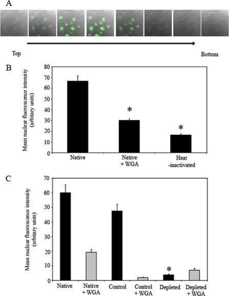
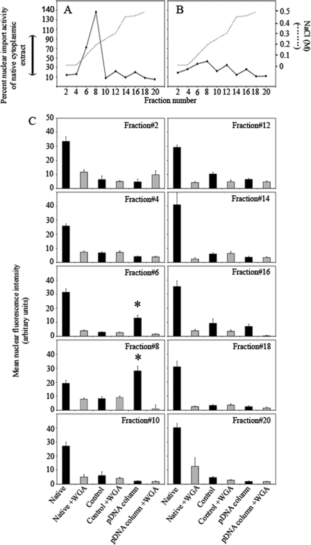
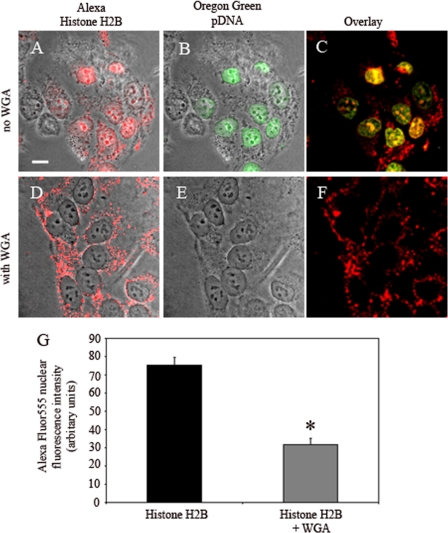
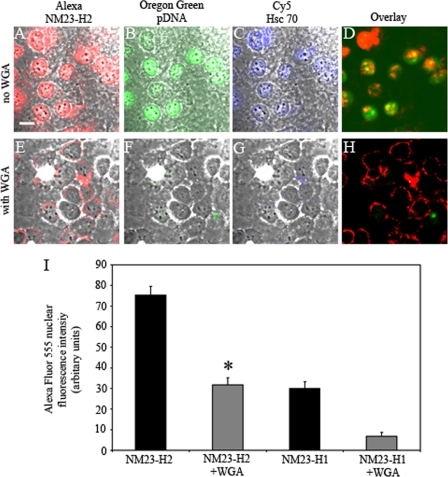
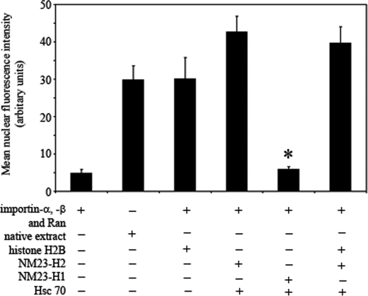
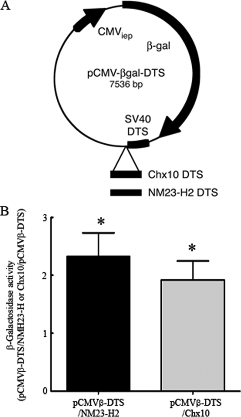
Import of exogenous plasmid DNA (pDNA) into mammalian cell nuclei represents a key intracellular obstacle to efficient non-viral gene delivery. This includes access of the pDNA to the nuclei of non-dividing cells where the presence of an intact nuclear membrane is limiting for gene transfer. Here we identify, isolate, and characterize, cytoplasmic determinants of pDNA nuclear import into digitonin-permeabilized HeLa cells. Depletion of putative DNA-binding proteins, on the basis of their ability to bind immobilized pDNA, abolished pDNA nuclear import supporting the critical role of cytoplasmic factors in this process. Elution of pDNA-bound proteins, followed by two-dimensional sodium dodecyl polyacrylamide gel electrophoresis identified several candidate DNA shuttle proteins. We show that two of these, NM23-H2, a ubiquitous c-Myc transcription-activating nucleoside diphosphate kinase, and the core histone H2B can both reconstitute pDNA nuclear import. Further, we demonstrate a significant increase in gene transfer in non-dividing HeLa cells transiently transfected with pDNA containing binding sequences from two of the DNA shuttle proteins, NM23-H2 and the homeobox transcription factor Chx10. These data support the hypothesis that exogenous pDNA binds to cytoplasmic shuttle proteins and is then translocated to the nucleus using the minimal import machinery. Importantly, increasing the binding of pDNA to shuttle proteins by re-engineering reporter plasmids with shuttle binding sequences enhances gene transfer. Increasing the potential for exogenously added pDNA to bind intracellular transport cofactors may enhance the potency of non-viral gene transfer.


Figures

FIGURE 1.
FIGURE 1.
Plasmid DNA nuclear import into digitonin-permeabilized HeLa cells. Digitonin-permeabilized HeLa cells were incubated in the presence of recombinant importin-α, -β, and Ran, with Oregon Green/pCMVβ-DTS (10 μg DNA/ml) and native cytoplasmic HeLa (0.5 mg protein/ml). A, confocal z-scan consisting of 8 optical sections (0.5-μm step size) superimposed on their corresponding phase contrast images from the top to the bottom of the nuclei to verify fluorescent pDNA nuclear localization (representative of six similar experiments). B, quantification of the nuclear fluorescence intensity in the central sections of images from the assay performed with native (in the absence or presence of WGA) or heat-inactivated extract. The bars represent mean ± S.E. of the background-subtracted nuclei fluorescent intensity (n = 24–35 nuclei). Cytoplasmic extract was heat-inactivated (90 °C, 10 min) before addition. Asterisks indicate a significant difference from native cytoplasmic extract import activity (p < 0.001). C, depletion of putative DNA-binding factors abolishes the plasmid DNA nuclear import activity from cytoplasmic extracts. The pDNA nuclear import activity of depleted HeLa cytoplasmic extract was compared with that of native and control extracts. Digitonin-permeabilized HeLa cells were incubated with the extracts (5 mg protein/ml) containing the Oregon Green/pCMVβ-DTS (10 μg/ml) in the absence (dark bars) or presence (gray bars) of WGA (100 μg/ml). After incubation, cells were processed for visualization by CLSM. Quantification of the nuclear fluorescence intensity in central sections is shown. 17–42 nuclei were analyzed for each experimental condition. The bars represent mean ± S.E. of the background-subtracted nuclei fluorescent intensity compared with native cytoplasmic extract in the absence of WGA (*, p < 0.001).
FIGURE 2.
FIGURE 2.
Elution profiles of HeLa cell cytoplasmic extracts on a pCMVβ-DTS/PNA-Sepharose column. Cytoplasmic extract (0.5 mg of protein in 500 μl) was applied to the pCMVβ-DTS/PNA-Sepharose column (A) or control PNA-Sepharose column (B), washed with three column volumes of column buffer, and elution achieved with a linear gradient of NaCl (0–0.5 m NaCl, per 20 ml buffer; 0.5 min/ml, 4 °C). Alternate elution fractions (2 ml each) were pooled (denoted numbers 2, 4, 6, 8, 10, 12, 14, 16, 18, and 20), dialyzed overnight against import buffer, concentrated using Centricon-5 microconcentration devices and assayed for Oregon Green/pCMVβ-DTS nuclear import activity. Quantification was performed on 8–59 nuclei for each type of treatment. Percent activity represents the mean background-subtracted nuclear fluorescent intensity as a percentage of the native HeLa cytoplasmic extract import activity. The concentration of the linear NaCl gradient is superimposed on the elution profile. C, fractionation of pDNA nuclear import activity from HeLa cell cytoplasmic extracts using the pCMVβ-DTS/PNA-Sepharose column. Details are as for A and B. Activity was determined in the absence (dark bars) or presence (gray bars) of WGA (100 μg/ml). Data points represent the mean nuclear fluorescence intensity ± S.E. (n = 8–59 nuclei).
FIGURE 3.
FIGURE 3.
Nuclear entry and plasmid DNA nuclear import activity of recombinant histone H2B. Digitonin-permeabilized HeLa cells were incubated with Oregon Green/pCMVβ-DTS (10 μg/ml), cytoplasmic extract (5 mg protein/ml), and Alexa histone H2B in the absence (A–C) or presence (D–F) of WGA (100 μg/ml). After incubation, cells were processed, as indicated under “Experimental Procedures” and fluorescence images of 0.5 μm optical sections and corresponding phase contrast images visualized using a CLSM. Representative central section fluorescent images matched and superimposed with their respective phase contrast images are shown. Alexa Fluor-555 (red) channel; (A, D), Oregon Green 488 (green) channel; (B, E). Alexa Fluor 555 confocal images for histone H2B were matched to their respective Oregon Green-pDNA images, and a picture overlay was performed using Adobe Photoshop to show co-localization in the absence (C) or presence of WGA (F). Bar, 10 μm. G, quantification of the Alexa-histone H2B nuclear fluorescence intensity in the central confocal sections. The bars represent mean ± S.E. of the background subtracted Oregon Green/pCMVβ-DTS nuclei fluorescence intensity (n = 33 nuclei). The asterisk indicates a significant (p < 0.001) difference from import activity supported by recombinant importin-α, -β, and Ran alone.
FIGURE 4.
FIGURE 4.
Nuclear entry and plasmid DNA nuclear import activity of recombinant NM23-H2. Digitonin-permeabilized HeLa cells were incubated with Oregon Green/pCMVβ-DTS, cytoplasmic extract, Cy5-Hsc70, and Alexa-NM23-H2 in the absence or presence of WGA. After incubation, cells were processed, as indicated under “Experimental Procedures,” and fluorescence images of 0.5-μm optical sections and corresponding phase contrast images visualized using a CLSM. Representative central section fluorescent images matched and superimposed with their respective phase contrast images are shown. Alexa-NM23-H2 in the absence (A–D) or presence (E–H) of WGA (100 μg/ml). Alexa Fluor-555 (red) channel; (A, E), Oregon Green 488 (green) channel; (B, F), Cy-5 (blue) channel; (C, G). Alexa Fluor 555 confocal images for NM23-H2 were matched to their respective Oregon Green-pDNA images and a picture overlay was performed to show co-localization in the absence (D) or presence of WGA (H). Bar, 10 μm. I, quantification of the Alexa-H2B and Hsc70-mediated Alexa-NM23-H2 nuclear fluorescence intensity in the central confocal sections. Bars represent mean ± S.E. of the background-subtracted Oregon Green/pCMVβ-DTS nuclei fluorescence intensity (n = 33–42 nuclei). The asterisk indicates significant (p < 0.001) difference from import activity supported by recombinant importin-α, -β, and Ran alone.
FIGURE 5.
FIGURE 5.
Quantification of the Oregon Green 488-labeled pDNA nuclear fluorescence intensity in the central confocal sections is shown. Bars represent mean ± S.E. of the mean background-subtracted Oregon Green/pCMVβ-DTS nuclei fluorescence intensity (n = 33–42 nuclei). The asterisk indicates significant (p < 0.001) difference from import activity supported by recombinant importin-α, -β, and Ran alone.
FIGURE 6.
FIGURE 6.
Plasmids (A). Abbreviated elements are as follows: CMViep, CMV immediate early promoter/enhancer; Chx10 or NM23-H2, binding sequences from shuttle proteins, Chx10 and NM23-H2, respectively; SV40 DTS, SV40 DNA nuclear targeting sequence. β-Galactosidase reporter gene activity of pCMVβ-DTS/NM23-H2 and pCMVβ-DTS/Chx10 (B). Forty-eight hours after Lipofectamine 2000-mediated transfection of G1/S-arrested HeLa cells with 1 μg of either control pCMVβ-DTS, pCMVβ-DTS/NM23-H2, or pCMVβ-DTS/Chx10, β-galactosidase activity was assayed. Data represent the mean ratio of the β-galactosidase activity (RLU/mg protein) in cells transfected with pCMVβ-DTS/NM23-H2 or pCMVβ-DTS/Chx10 and pCMVβ-DTS (means ± S.E. error bars; n = 12 dishes from three independent experiments).

Similar articles

Cited by

References

    1. Nishikawa M., Huang L. (2001) Hum. Gene Ther. 12, 861–870 - PubMed
    1. Görlich. D., Kutay U. (1999) Annu. Rev. Cell. Dev. Biol. 15, 607–660 - PubMed
    1. Tseng W. C., Haselton F. R., Giorgio T. D. (1999) Biochim. Biophys. Acta 1445, 53–64 - PubMed
    1. Wilson G. L., Dean B. S., Wang G., Dean D. A. (1999) J. Biol. Chem. 274, 22025–22032 - PMC - PubMed
    1. Cartier R., Reszka R. (2002) Gene Therapy 9, 157–167 - PubMed

Publication types

MeSH terms