. 2009 Sep 25;284(39):26591-602.
doi: 10.1074/jbc.M109.022491. Epub 2009 Jul 28.

JNK1-dependent PUMA expression contributes to hepatocyte lipoapoptosis

Affiliations

JNK1-dependent PUMA expression contributes to hepatocyte lipoapoptosis

Sophie C Cazanave et al. J Biol Chem. .

Abstract

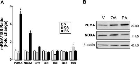
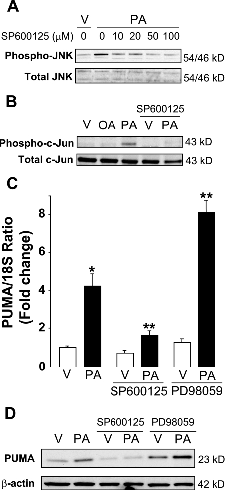
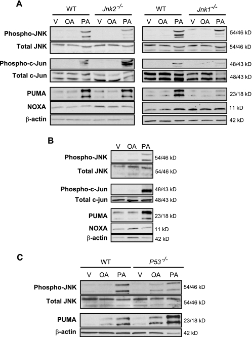
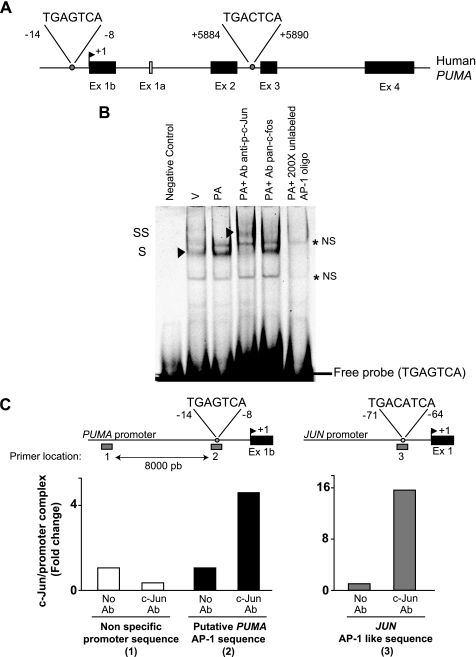
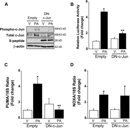
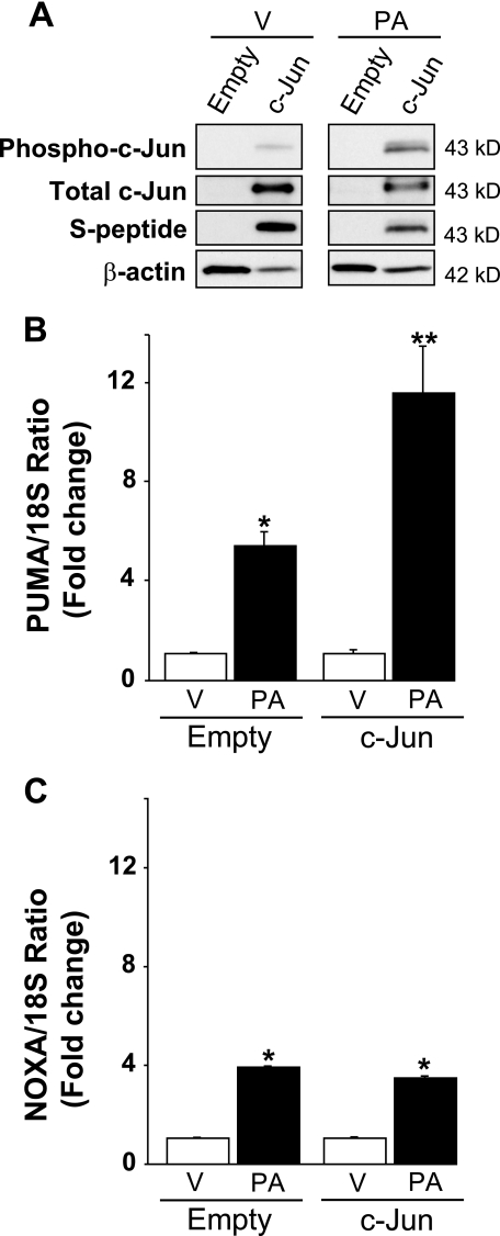
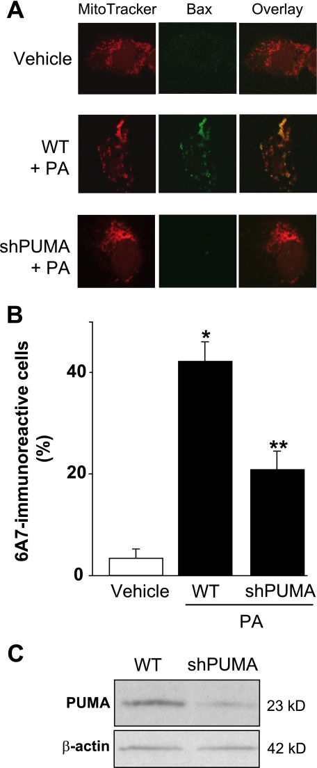
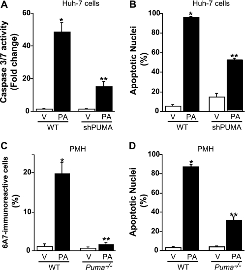
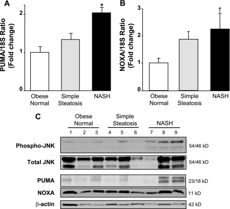
Free fatty acids (FFA) induce hepatocyte lipoapoptosis by a c-Jun N-terminal kinase (JNK)-dependent mechanism. However, the cellular processes by which JNK engages the core apoptotic machinery during lipotoxicity, especially activation of BH3-only proteins, remain incompletely understood. Thus, our aim was to determine whether JNK mediates induction of BH3-only proteins during hepatocyte lipoapoptosis. The saturated FFA palmitate, but not the monounsaturated FFA oleate, induces an increase in PUMA mRNA and protein levels. Palmitate induction of PUMA was JNK1-dependent in primary murine hepatocytes. Palmitate-mediated PUMA expression was inhibited by a dominant negative c-Jun, and direct binding of a phosphorylated c-Jun containing the activator protein 1 complex to the PUMA promoter was identified by electrophoretic mobility shift assay and a chromatin immunoprecipitation assay. Short hairpin RNA-targeted knockdown of PUMA attenuated Bax activation, caspase 3/7 activity, and cell death. Similarly, the genetic deficiency of Puma rendered murine hepatocytes resistant to lipoapoptosis. PUMA expression was also increased in liver biopsy specimens from patients with non-alcoholic steatohepatitis as compared with patients with simple steatosis or controls. Collectively, the data implicate JNK1-dependent PUMA expression as a mechanism contributing to hepatocyte lipoapoptosis.


Figures

FIGURE 1.
FIGURE 1.
Palmitate increases PUMA mRNA and protein levels. Total RNA and whole cell lysates were prepared from Huh-7 cells treated with palmitic acid (PA), oleic acid (OA) at 800 μm, and vehicle (V) was used as control. A, BH3-only protein mRNA expression was profiled by real-time PCR 8 h after FFA treatment. -Fold induction was determined after normalization to 18 S. Data represent the mean and S.E. of four independent experiments; *, p < 0.01, palmitate-treated cells versus vehicle- or oleate-treated cells. B, immunoblot analysis was performed for PUMA and NOXA protein expression 16 h after treatment with FFA. β-Actin was used as control for protein loading.
FIGURE 2.
FIGURE 2.
JNK pharmacological inhibition prevents palmitate-induced PUMA expression. A, whole cell lysates were prepared from Huh-7 cells treated with palmitic acid (PA) at 800 μm in the presence of serial dose of SP600125 (10–100 μm) for 2 h. Vehicle (V) was used as control. Immunoblot analysis was performed for phosphorylated JNK protein expression and total JNK, a control for protein loading. B, whole cell lysates were prepared from Huh-7 cells treated with vehicle (V), palmitic acid (PA), or oleic acid (OA) at 800 μm and from Huh-7 cells treated with vehicle or palmitic acid in the presence of SP600125 (50 μm). Immunoblot analysis was performed for phosphorylated c-Jun protein expression 16 h after treatment with FFA. Total c-Jun was used as a control for protein loading. C, total RNA were prepared from Huh-7 cells treated with vehicle or palmitic acid (800 μm) in the presence of SP600125 (50 μm) or PD98059 (50 μm) for 8 h. PUMA mRNA expression was profiled by real time PCR. -Fold induction was determined after normalization to 18 S. Data represent the mean and S.E. of three independent experiments. *, p < 0.01, palmitate-treated cells versus vehicle-treated cells. **, p < 0.01, palmitate-treated cells in the presence of inhibitors versus palmitate-treated cells. D, whole cell lysates were prepared from Huh-7 cells treated with vehicle or palmitic acid (800 μm) in the presence of SP600125 (50 μm) or PD98059 (50 μm) for 16 h. Immunoblot analysis was performed for PUMA protein expression, and β-actin was used as control for protein loading.
FIGURE 3.
FIGURE 3.
JNK1-mediated c-Jun phosphorylation induces PUMA expression in primary hepatocytes during lipoapoptosis. Whole cell lysates were obtained from isolated primary mouse and human hepatocytes treated with palmitic acid (PA) or oleic acid (OA) at 800 μm for 8 h. Vehicle (V) was used as control. Immunoblot analysis was performed for phosphorylated and total JNK, phosphorylated and total c-Jun, PUMA, NOXA, and β-actin. A, primary murine hepatocytes isolated from WT, Jnk1−/−, and Jnk2−/− mice. B, primary human hepatocytes. C, primary murine hepatocytes isolated from WT and p53−/− mice.
FIGURE 4.
FIGURE 4.
Palmitate-induced AP-1 complex binds to the PUMA promoter. A, the genomic structure of PUMA showing the exon-intron organization and 2 potential AP-1 binding sites. B, nuclear protein extracts isolated from Huh-7 cells treated with vehicle (V) or palmitic acid (PA) at 800 μm for 12 h. EMSA was performed using CY 5.5-labeled double-stranded oligonucleotide containing the putative AP-1 binding sequence within the human PUMA promoter (located at −14/−8 nucleotides from the transcriptional start codon), supershift experiments using antibody against phospho-c-Jun or pan-c-Fos, and competition experiments with 200-fold molar excess cold oligonucleotide as described under “Experimental Procedures.” Retarded complexes are indicated by arrows (S, shift; SS, supershift; NS, nonspecific band). C, Huh-7 cells were treated with palmitic acid (800 μm) for 2 h. Next, the cells were fixed and lysed, and DNA fragments co-immunoprecipitated with the target protein c-Jun were subjected to quantitative real-time PCR analysis using various primer sets as indicated by the gray bars. The primer set 1 (P1) and 2 (P2) were used to amplify, respectively, an upstream nonspecific sequence or the putative AP-1 sequence within the human PUMA promoter; the primer set 3 (P3) was used to amplify the AP-1 like sequence in the human JUN promoter. As a control, a mock immunoprecipitation without antibody (Ab) was also performed. Representative results of three independent experiments are shown.
FIGURE 5.
FIGURE 5.
Enforced expression of dominant negative c-Jun decreased phosphorylation of endogenous c-Jun and AP-1 transcriptional activity and prevents induction of PUMA mRNA level in response to palmitate. Huh-7 cells were transiently transfected for 24 h with an S-peptide-tagged dominant negative c-Jun encoding plasmid (DN-c-Jun) or the control empty plasmid. Next, cells were treated with palmitic acid (PA) at 800 μm. Vehicle (V) was used as control. A, whole cell lysates were prepared 8 h after FFA treatment, and effective expression of dominant negative c-Jun was identified by immunoblot analysis using an anti-S-peptide antibody (which reveals a band at approximately 28 kDa). Immunoblot analysis was also performed for phosphorylated and total c-Jun and β-actin. B, using pAP1-Luc vector, relative luciferase activity was assessed 24 h after FFA treatment in the presence of the pan-caspase inhibitor benzyloxycarbonyl-Val-Ala-Asp-fluoromethyl ketone (50 μm) to mitigate cell death, as described under “Experimental Procedures.” Data represent the mean and S.E. of three independent experiments; *, p < 0.01, palmitate-treated empty vector versus vehicle-treated empty vector; **, p < 0.01, palmitate-treated DN-c-Jun versus palmitate-treated empty vector. Total RNA was extracted 8 h after FFA treatment, and PUMA (C) and NOXA (D) mRNA expression was profiled by real-time PCR. -Fold induction was determined after normalization to 18 S. Data represent the mean and S.E. of three experiments; *, p < 0.01, palmitate-treated cells versus vehicle-treated cells; **, p < 0.01, palmitate-treated DN-c-Jun versus palmitate-treated empty vector.
FIGURE 6.
FIGURE 6.
Enhanced expression of c-Jun exacerbates palmitate induction of PUMA mRNA levels. Huh-7 cells were transiently transfected for 24 h with a plasmid containing full-length of human c-Jun open reading frame tagged with S-peptide (c-Jun) or the empty vector. Total RNA and whole cell lysates were prepared 8 h after treatment with palmitic acid (PA) at 800 μm, and vehicle (V) was used as the control. A, effective overexpression of c-Jun protein level was verified by immunoblot analysis using an anti-S-peptide antibody and an anti-total c-Jun antibody. Immunoblot analysis was also performed for phosphorylated c-Jun and β-actin. PUMA (B) and NOXA (C) mRNA expression was profiled by real-time PCR. -Fold induction was determined after normalization to 18 S. Data represent the mean and S.E. of three experiments. *, p < 0.05, palmitate-treated cells versus vehicle-treated cells; **, p < 0.01, palmitate-treated c-Jun versus palmitate-treated empty vector.
FIGURE 7.
FIGURE 7.
PUMA contributes to Bax activation. A and B, Huh-7 (WT) and Huh-7 cells stably expressing short hairpin RNA complementary to PUMA (shPUMA) were incubated for 12 h with palmitic acid (PA) at 800 μm; vehicle (V)-treated cells were used as control. Cells were stained with Mito Tracker Red to visualize mitochondria. Next, cells were fixed, and Bax activation was assessed using conformation specific antisera (6A7), which only recognizes active Bax and using immunofluorescence microscopy. A, representative images of three independent experiments are depicted. B, 6A7-immunoreactive cells were quantified in 10 random 40× objective fields for each condition at the indicated time point with automated software. Data represent the mean and S.E. of three experiments; *, p < 0.01, palmitate-treated cells versus vehicle-treated cells; **, p < 0.01, palmitate-treated shPUMA versus palmitate-treated WT. C, effective down-regulation of PUMA protein levels in shPUMA Huh-7 cells compared with WT Huh-7 cells was verified by immunoblot analysis on whole cell lysates.
FIGURE 8.
FIGURE 8.
PUMA contributes to palmitate-mediated apoptosis. A and B, Huh-7 (WT) and Huh-7 cells stably expressing short hairpin RNA complementary to PUMA (shPUMA) were incubated for 24 h with palmitic acid (PA) at 800 μm; Vehicle (V)-treated cells were used as control. A, caspases 3 and 7 catalytic activity was assessed as described under “Experimental Procedures.” B, apoptotic nuclei were counted according to morphological criteria after 4′,6-diamidino-2-phenylindole dihydrochloride staining. Data represent the mean and S.E. of three experiments; *, p < 0.05, palmitate-treated cells versus vehicle-treated cells; **, p < 0.01, palmitate-treated shPUMA versus palmitate-treated WT. C, primary murine hepatocytes (PMH) isolated from WT or Puma−/− mice were treated for 8 h with vehicle or palmitic acid (400 μm). Bax activation was assessed as described under “Experimental Procedures,” and 6A7-immunoreactive cells were quantified in 12 random 40× objective fields for each condition with automated software. Data represent the mean and S.E. of three experiments; *, p < 0.01, palmitate-treated cells versus vehicle-treated cells; **, p < 0.01, palmitate-treated Puma−/− versus palmitate-treated WT. D, primary murine hepatocytes isolated from WT or Puma−/− mice were treated for 24 h with vehicle or palmitic acid (200 μm). Apoptotic nuclei were counted according to morphological criteria after 4′,6-diamidino-2-phenylindole dihydrochloride staining. Data represent mean and S.E. of three experiments; *, p < 0.01, palmitate-treated cells versus vehicle-treated cells; **, p < 0.01, palmitate-treated Puma−/− versus palmitate-treated WT.
FIGURE 9.
FIGURE 9.
PUMA is up-regulated in human NASH. Homogenates and total RNA were prepared from human liver biopsies of obese normal (n = 16), simple steatosis (n = 17), or NASH patients (n = 16). A and B, PUMA and NOXA mRNA expression was profiled by real-time PCR. -Fold induction was determined after normalization to 18 S. Data represent the mean and S.E.; *, p < 0.01, NASH patients versus obese normal and simple steatosis patients; , p < 0.05, NASH patient versus obese normal patients. C, immunoblot analysis was performed for phosphorylated and total JNK, phosphorylated and total c-Jun, PUMA, NOXA, and β-actin.

References

    1. Browning J. D., Szczepaniak L. S., Dobbins R., Nuremberg P., Horton J. D., Cohen J. C., Grundy S. M., Hobbs H. H. (2004) Hepatology 40, 1387–1395 - PubMed
    1. Adams L. A., Lymp J. F., St Sauver J., Sanderson S. O., Lindor K. D., Feldstein A., Angulo P. (2005) Gastroenterology 129, 113–121 - PubMed
    1. Ekstedt M., Franzén L. E., Mathiesen U. L., Thorelius L., Holmqvist M., Bodemar G., Kechagias S. (2006) Hepatology 44, 865–873 - PubMed
    1. Ratziu V., Poynard T. (2006) Hepatology 44, 802–805 - PubMed
    1. Bookman I. D., Pham J., Guindi M., Heathcote E. J. (2006) Liver Int. 26, 566–571 - PubMed

Publication types

MeSH terms