. 2009 Nov;23(11):3917-27.
doi: 10.1096/fj.09-136630. Epub 2009 Jul 28.

SUMOylation of the mitochondrial fission protein Drp1 occurs at multiple nonconsensus sites within the B domain and is linked to its activity cycle

Affiliations

SUMOylation of the mitochondrial fission protein Drp1 occurs at multiple nonconsensus sites within the B domain and is linked to its activity cycle

Claudia Figueroa-Romero et al. FASEB J. 2009 Nov.

Abstract

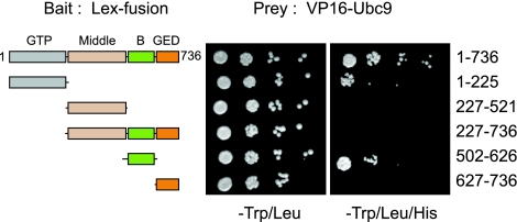
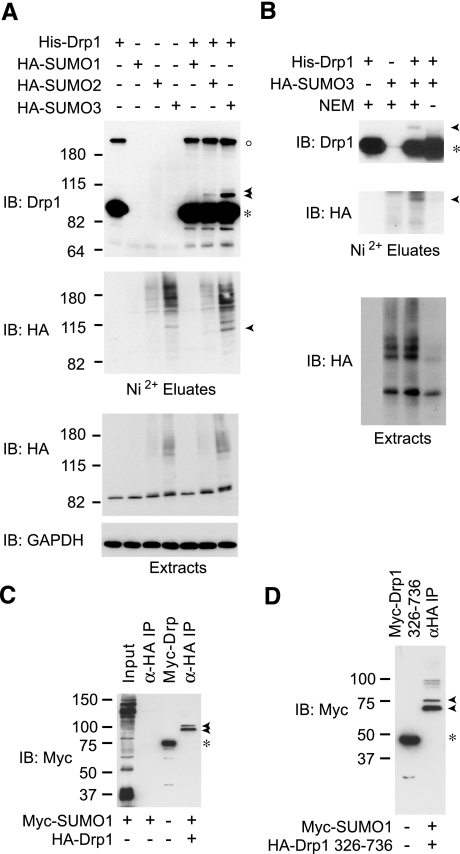
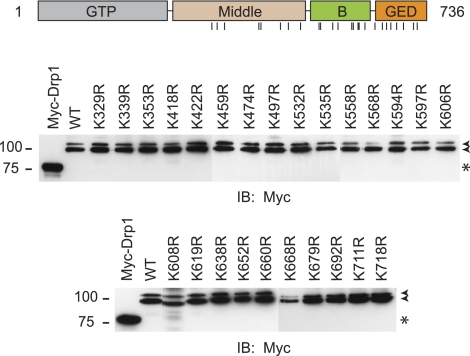
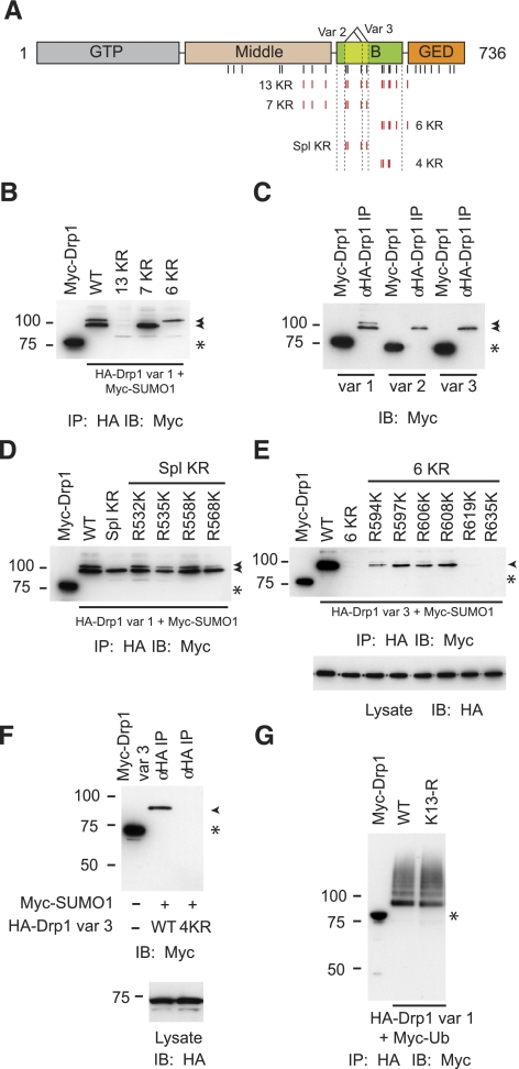
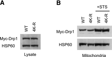
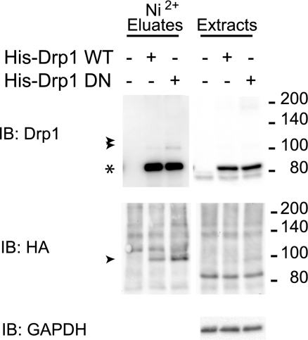
Dynamin-related protein (Drp) 1 is a key regulator of mitochondrial fission and is composed of GTP-binding, Middle, insert B, and C-terminal GTPase effector (GED) domains. Drp1 associates with mitochondrial fission sites and promotes membrane constriction through its intrinsic GTPase activity. The mechanisms that regulate Drp1 activity remain poorly understood but are likely to involve reversible post-translational modifications, such as conjugation of small ubiquitin-like modifier (SUMO) proteins. Through a detailed analysis, we find that Drp1 interacts with the SUMO-conjugating enzyme Ubc9 via multiple regions and demonstrate that Drp1 is a direct target of SUMO modification by all three SUMO isoforms. While Drp1 does not harbor consensus SUMOylation sequences, our analysis identified2 clusters of lysine residues within the B domain that serve as noncanonical conjugation sites. Although initial analysis indicates that mitochondrial recruitment of ectopically expressed Drp1 in response to staurosporine is unaffected by loss of SUMOylation, we find that Drp1 SUMOylation is enhanced in the context of the K38A mutation. This dominant-negative mutant, which is deficient in GTP binding and hydrolysis, does not associate with mitochondria and prevents normal mitochondrial fission. This finding suggests that SUMOylation of Drp1 is linked to its activity cycle and is influenced by Drp1 localization.


Figures

Figure 1.
Figure 1.
Ubc9 interacts with the GTPase and B domains of Drp1 in the yeast 2-hybrid assay. L40 yeast cells were cotransformed with vectors expressing fusion proteins of LexA DNA binding domain (LexA) (bait) with full-length Drp1 (aa 1–736), GTPase (GTP) domain (aa 1–225), Middle domain (aa 227–521), no-GTPase domain (aa 227–736), B domain (aa 502–626), or GTPase-effector (GED) domain (aa 627–736) and vectors expressing Ubc9 as a fusion protein with the VP16 activation domain (VP16) (prey). Cotransformed colonies were grown on −Trp/Leu medium; serial yeast dilutions were plated. Expression of both plasmids is confirmed by growth in −Trp/Leu plates; interaction between proteins was assessed by growth of yeast on minimal medium plates (−Trp/Leu/His),indicating activation of the HIS reporter. None of the baits conferred histidine auxotrophy in the absence of the Ubc9 prey (data not shown).
Figure 2.
Figure 2.
Drp1 is modified by SUMO1, SUMO2, and SUMO3. A) HEK-293 cells were transfected transiently with expression vectors for pcDNA3-Ubc9 and/or pcDNA3-HA-SUMO1, pcDNA3-HA-SUMO2, or pcDNA3-HA-SUMO3 and/or pcDNA3-V5-His-Drp1. Cells were lysed in the presence of 20 mM NEM, and His-Drp1 was isolated via Ni2+ chelate chromatography under denaturing conditions. Proteins from Ni2+ eluates and extracts were visualized by Western immunoblot (IB) analysis using antibodies against Drp1, HA-epitope, and GAPDH. Migrations of unmodified and SUMO-conjugated forms of Drp1 are indicated by asterisk and arrowheads, respectively. Open circle indicates larger apparent molecular mass Drp1 forms likely representing entangled SDS-resistant oligomers. B) SUMO modification of Drp1 is Ubc9 and SUMO3 dependent and NEM sensitive. HEK-293 cells transiently transfected with pcDNA3-Ubc9 and pcDNA3-HA-SUMO3 and/or pcDNA3-V5-His-Drp1 were lysed in the presence or absence of 20 mM NEM and analyzed as in A. C) Cells were cotransfected with HA-Drp1 and Myc-SUMO1, or else transfected with Myc-SUMO1 alone, and cell lysates were immunoprecipitated using anti-HA antibodies as indicated (α-HA IP), then immunoblotted using anti-Myc antibodies. Arrowheads indicate SUMO-modified Drp1 forms. Myc-Drp1 expression (asterisk) identifies the size of unmodified Drp1, as it is identical in size to HA-Drp1. D) Cells were cotransfected with HA-Drp1 (aa 326–736) and Myc-SUMO1, and extracts were immunoprecipitated with anti-HA antibodies and then immunoblotted for Myc as in C. Arrowheads indicate SUMO-modified Drp1 (aa 326–736) forms. Myc-Drp1 (aa 326–736) expression (asterisk) identifies the size of unmodified Drp1, which is identical in size to HA-Drp1 (aa 326–736). Migrations of molecular mass standards (kDa) are at left.
Figure 3.
Figure 3.
Mutational analysis of potential SUMO-acceptor lysines. Schematic diagram of Drp1 domain organization (top panel); vertical lines identify lysine residues within aa 326–736 mutated to arginines in the bottom panels. Cells were cotransfected with Myc-SUMO1 and WT HA-Drp1 (aa 1–736; splice variant 1) or the indicated Drp1 mutants (single amino acid letter code). Extracts were immunoprecipitated with anti-HA antibodies and then immunoblotted for Myc. Arrowheads indicate SUMO-modified Drp1. Myc-Drp1 expression (asterisk) identifies the size of unmodified HA-Drp1. Migrations of molecular mass standards (kDa) are at left.
Figure 4.
Figure 4.
Noncanonical SUMOylation sites are present within the B domain of Drp1. A) Schematic diagram of Drp1; vertical lines identify lysine residues mutated to arginines in the other panels. In the 5 constructs at bottom, indicated Lys residues (red) were converted to Arg residues, and constructs were named as shown. B) Cells were cotransfected with Myc-SUMO1 and WT HA-Drp1 (aa 1–736; splice variant 1) or the indicated Drp1 mutants shown in A. Extracts were immunoprecipitated with anti-HA antibodies and then immunoblotted for Myc. Arrowheads indicate SUMO-modified Drp1, as in subsequent panels. Myc-Drp1 expression (asterisk) identifies size of unmodified HA-Drp1, as in subsequent panels. C) Cells were transfected and analyzed as in B, examining the 3 splice variants of WT Drp1. D) Cells were transfected and analyzed as in B, with indicated mutated residues in Drp1 Spl KR individually returned to the Lys residues found in the WT form. E) Cells were transfected and analyzed as in D, though HA-Drp1 was the variant 3 form, and the residues were reverted from the 6 KR mutant. Bottom panel: total cell lysates were immunoblotted for HA to demonstrate equal expression of all Drp1 forms. F) Cells were cotransfected with Myc-SUMO1 and either WT or 4 KR mutant HA-Drp1 (splice variant 3). Extracts were immunoprecipitated with anti-HA antibodies and then immunoblotted for Myc (top panel) or HA (bottom panel). G) Cells were cotransfected with Myc-ubiquitin and either WT or 13 KR mutant HA-Drp1 (splice variant 1). Extracts were immunoprecipitated with anti-HA antibodies and then immunoblotted for Myc.
Figure 5.
Figure 5.
Preventing Drp1 SUMOylation does not alter its translocation to mitochondria. HeLa cells were transfected with Myc-tagged, WT Drp1 variant 3 or the 4 KR mutant form that cannot be SUMOylated. A) Total cell lysates were immunoblotted for Myc. HSP60 levels were monitored on the same immunoblot to ensure equal protein loading. B) Cells expressing WT or 4 KR mutant Drp1 were either left untreated or else treated with staurosporine (STS) in the presence of zVAD-fmk for 8 h. Mitochondria were isolated; equal protein aliquots were resolved by SDS-PAGE, then immunoblotted for Myc and HSP60.
Figure 6.
Figure 6.
Enhancement of SUMO modification of dominant-negative Drp1. HEK-293 cells were transiently transfected with pcDNA3-V5-Ubc9, pcDNA3-HA-SUMO3, and/or pcDNA3-V5-His-Drp1 WT or pcDNA3-V5-His-Drp1 K38A, and CS2+empty vector. Protein extracts were obtained in the presence of 20 mM NEM, processed as in Fig. 2A, and analyzed by immunoblotting. Arrowheads indicate modified doublet; asterisk indicates unmodified Drp1.

Similar articles

Cited by

References

    1. Cerveny K L, Tamura Y, Zhang Z, Jensen R E, Sesaki H. Regulation of mitochondrial fusion and division. Trends Cell Biol. 2007;17:563–569. - PubMed
    1. Griparic L, van der Wel N N, Orozco I J, Peters P J, van der Bliek A M. Loss of the intermembrane space protein Mgm1/OPA1 induces swelling and localized constrictions along the lengths of mitochondria. J Biol Chem. 2004;279:18792–18798. - PubMed
    1. Santel A, Frank S, Gaume B, Herrler M, Youle R J, Fuller M T. Mitofusin-1 protein is a generally expressed mediator of mitochondrial fusion in mammalian cells. J Cell Sci. 2003;116:2763–2774. - PubMed
    1. Wells R C, Picton L K, Williams S C, Tan F J, Hill R B. Direct binding of the dynamin-like GTPase, Dnm1, to mitochondrial dynamics protein Fis1 is negatively regulated by the Fis1 N-terminal arm. J Biol Chem. 2007;282:33769–33775. - PMC - PubMed
    1. Yoon Y, Krueger E W, Oswald B J, McNiven M A. The mitochondrial protein hFis1 regulates mitochondrial fission in mammalian cells through an interaction with the dynamin-like protein DLP1. Mol Cell Biol. 2003;23:5409–5420. - PMC - PubMed

Publication types

MeSH terms

Substances