. 2009 Aug 15;122(Pt 16):2914-23.
doi: 10.1242/jcs.046789. Epub 2009 Jul 28.

The dynamic properties of intermediate filaments during organelle transport

Affiliations

The dynamic properties of intermediate filaments during organelle transport

Lynne Chang et al. J Cell Sci. .

Abstract

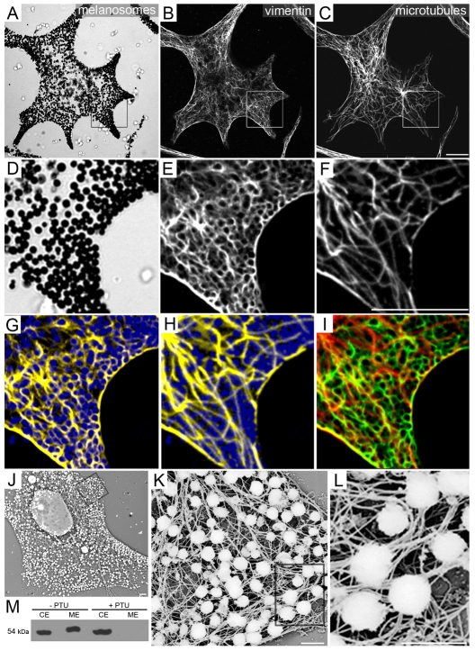
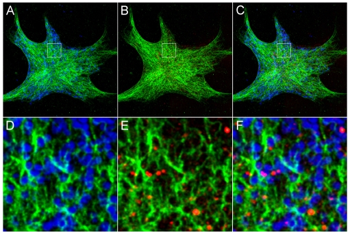
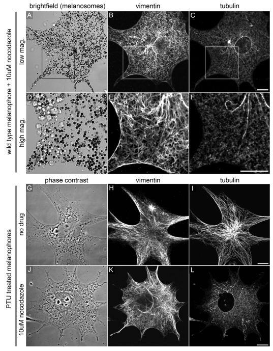
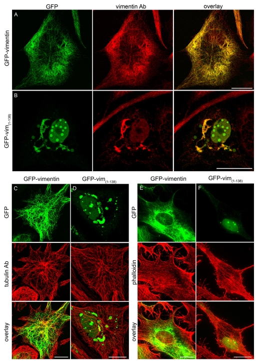
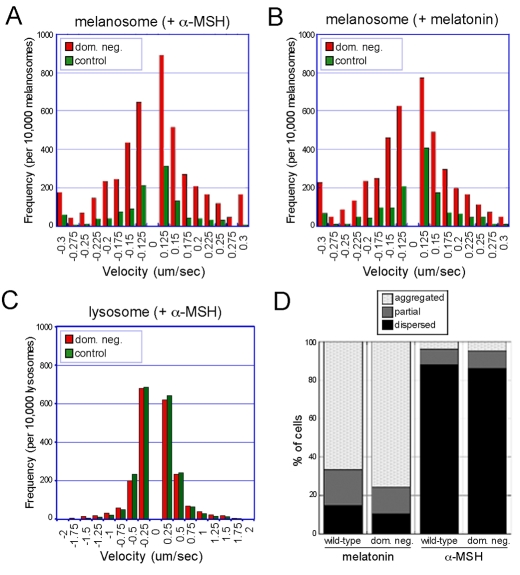
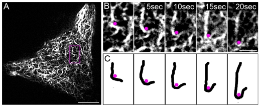
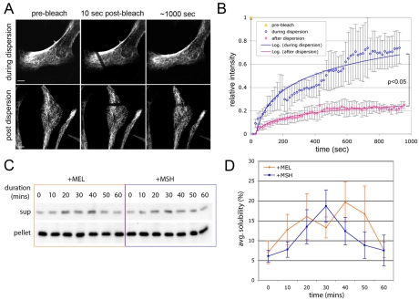
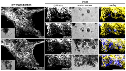
Intermediate filament (IF) dynamics during organelle transport and their role in organelle movement were studied using Xenopus laevis melanophores. In these cells, pigment granules (melanosomes) move along microtubules and microfilaments, toward and away from the cell periphery in response to alpha-melanocyte stimulating hormone (alpha-MSH) and melatonin, respectively. In this study we show that melanophores possess a complex network of vimentin IFs which interact with melanosomes. IFs form an intricate, honeycomb-like network that form cages surrounding individual and small clusters of melanosomes, both when they are aggregated and dispersed. Purified melanosome preparations contain a substantial amount of vimentin, suggesting that melanosomes bind to IFs. Analyses of individual melanosome movements in cells with disrupted IF networks show increased movement of granules in both anterograde and retrograde directions, further supporting the notion of a melanosome-IF interaction. Live imaging reveals that IFs, in turn, become highly flexible as melanosomes disperse in response to alpha-MSH. During the height of dispersion there is a marked increase in the rate of fluorescence recovery after photobleaching of GFP-vimentin IFs and an increase in vimentin solubility. These results reveal a dynamic interaction between membrane bound pigment granules and IFs and suggest a role for IFs as modulators of granule movement.


Figures

Fig. 1.
Fig. 1.
Microscopic and biochemical evidence for vimentin association with melanosomes in Xenopus melanophores. (A-C) Immunofluorescence preparation of melanophores reveals a dense network of vimentin filaments (B) that permeate the cytoplasm. Microtubules form a sparse, radial network (C). Scale bar: 5 μm. (D-F) Higher magnification images of boxed regions in A-C show melanosomes (D) surrounded by a dense network of vimentin filaments (E). Scale bar: 5 μm. (G-I) Pseudo-colored overlays of D-F. Vimentin fibers (yellow) wrap around individual and clusters of melanosomes (blue; G). This encaging is not observed between microtubules (yellow) and melanosomes (blue; H). An overlay of vimentin (green) and microtubules (red) is shown in I. (J-L) Melanophores were processed for platinum replica electron microscopy using a technique that preserves intermediate filaments and dissolves microtubules (MTs) and actin filaments (MFs) (Svitkina et al., 1995) (see Materials and Methods). Melanosomes appear to remain closely associated with vimentin filaments in the absence of MTs and MFs. K and L are higher magnification views of J. Scale bars: (J) 2 μm; (K,L) 1 μm. (M) Western blot analysis of purified melanosomes using an antibody specific for vertebrate vimentin shows co-purification of vimentin with the granules in wild-type cells (–PTU; CE, total cell extract; ME, melanosome extract). Vimentin co-purifies with granules isolated from melanophores treated with either melatonin or α-melanocyte stimulating hormone (α-MSH; unpublished data). In PTU-treated cells that lack melanosomes, vimentin is not present in the pellet (+PTU).
Fig. 2.
Fig. 2.
Lysosomes are not closely associated with vimentin IFs. Melanophores were incubated with LysoTracker Red for 1 hour to visualize lysosomes, followed by processing for immunofluorescence to detect vimentin. Melanosomes were imaged using bright-field illumination and the image was inverted and given a blue hue in Photoshop. A-C show a low magnification, double (A,B) and triple (C) overlay images of vimentin (green), melanosomes (blue) and fluorescently labeled lysosomes (red). (D-F) Higher magnification images of the boxed regions in A-C show very few lysosomes that are closely associated with vimentin IFs.
Fig. 3.
Fig. 3.
Vimentin IFs association with melanosomes is independent of microtubules. (A-C) The distribution of vimentin IFs is not affected by nocodazole-induced depolymerization of MTs. The IF network remains fully extended and densely distributed throughout the cytoplasm. See Fig. 1A-F for comparison. Scale bar: 5 μm. (D-F) Higher magnification images of the boxed regions in A-C show vimentin filaments surrounding melanosomes in the absence of MTs. Scale bar: 5 μm. (G-I) The vimentin IF network is similarly distributed as an extended network in PTU-treated melanophores that lack melanosomes. Scale bar: 5 μm. (J-L) Nocodazole treatment of these cells does not appear to cause reorganization of the vimentin IF network, demonstrating that vimentin IF distribution is not a result of melanosomes anchoring IFs. Scale bar: 5 μm.
Fig. 4.
Fig. 4.
GFP-vimentin incorporates into the endogenous vimentin IF network and a GFP-tagged truncated vimentin construct disrupts the endogenous network in Xenopus melanophores. (A) Cells were transfected with a GFP-tagged full-length Xenopus vimentin construct for 48 hours. The GFP-vimentin (shown in green) co-assembles with endogenous vimentin (visualized by antibody staining; shown in red). (B) Cells transfected for 48 hours with a dominant-negative construct containing the head and alpha-helical domain 1A of vimentin [GFP-vim(1-138)] have a disrupted endogenous network. The truncated vimentin forms aggregates in the nucleus and perinuclear area, and sequesters the endogenous vimentin away from the peripheral cytoplasm. The truncated vimentin was tagged with a C-terminal GFP (shown in green), and the endogenous network was visualized by immunostaining (shown in red). (C,D) Disruption of vimentin IFs does not affect the distribution of microtubules. Cells were transfected with either full-length GFP-vimentin (C; green) or GFP-vim(1-138) (D; green) for 48 hours and immunostained with a microtubule-specific antibody (red). (E,F) Disruption of vimentin IFs does not affect the distribution of actin filaments. Cells were transfected with either full-length GFP-vimentin (E; green) or GFP-vim(1-138) (F; green) for 48 hours. Actin filaments were visualized by phalloidin (red). Scale bars: 20 μm.
Fig. 5.
Fig. 5.
Melanosomes move more in the absence of a vimentin IF network. (A,B) The number of moving melanosomes and their speed were elevated in both retrograde (–) and anterograde (+) directions in cells expressing the dominant negative vimentin construct, GFP-vim(1-138) (dom. neg.), when compared with control cells. These increases were evident during both dispersion by α-MSH treatment and melatonin-induced aggregation. (C) In contrast to melanosomes, lysosomal movement was not affected by the expression of GFP-vim(1-138) (dom. neg.) as compared with control cells. Lysosomes were tracked in cells treated with α-MSH to mirror the melanosome dispersion situation. Similar results were obtained in unstimulated cells (not shown). (D) Disruption of the vimentin IF network by overexpression of the dominant negative vimentin (dom. neg.), did not affect the end states of melanosomes in response to melatonin and α-MSH at 60 minutes after hormone treatment. Melanosomes were predominantly aggregated in both transfected and wild-type cells in response to melatonin and dispersed in both cell types in response to α-MSH.
Fig. 6.
Fig. 6.
Vimentin filaments undergo dramatic shape changes during melanosome movement. Xenopus melanophores expressing GFP-vimentin were used for live-imaging experiments. These cells were serum starved for 2 hours, followed by treatment with α-MSH to stimulate granule dispersion and then imaged using a spinning disk confocal microscope with 5-second frame intervals. (A) A low magnification image at time point 0 (scale bar: 5 μm). (B) Higher magnification, time-lapse frames show a granule, pseudo-colored purple, moving with the vimentin filament that bends and moves toward the lower left field of view (scale bar: 1 μm; also see supplementary material Movie 1). (C) A cartoon depiction of the relevant filament and granule from B, for easier visualization.
Fig. 7.
Fig. 7.
There is a higher fraction of exchangeable and soluble vimentin during hormone-induced melanosome movement than in steady state cells. (A) GFP-vimentin-expressing Xenopus melanophores (stable cell line) were imaged before and during recovery after photobleaching. The fluorescence recovery of cells (treated with α-MSH) during active melanosome dispersion and post-dispersion was compared. Contrast was adjusted in the 1000 second frame to visually normalize for overall loss of fluorescence during imaging. Scale bars: 10 μm. (B) Quantitative analysis of fluorescence recovery after photobleaching, both during (blue) and after (pink) melanosome dispersion. The result suggests a higher fraction of exchangeable vimentin during melanosome dispersion. The values are averages from four cells under each condition. The difference between the average t1/2s between dispersing and fully dispersed cells was statistically significant (P=0.037). Error bars indicate the standard deviation. (C) Western blot analysis shows a transient increase in vimentin solubility in response to melatonin (+MEL) or α-MSH (+MSH) treatment, followed by a decrease to initial levels. Soluble (sup) and pelletable fractions (pellet) from IF-enriched preparations were separated by centrifugation at 12,000 g for 10 minutes (see Materials and Methods). (D) Changes in the percentage of soluble vimentin in response to hormone treatment were quantified from western blots. Averages from three experiments for each treatment are shown (blue, α-MSH; orange, melatonin). The increase in solubility from time 0 to 40 minutes after treatment with melatonin was statistically significant (P=0.02). Similarly, changes in solubility between 0 and 30 minutes after treatment with α-MSH were also statistically significant (P=0.0072). The decrease in solubility between 40 or 30 minutes and 60 minutes after treatment was also significant for both treatments (P=0.018 for melatonin and 0.026 for α-MSH). Error bars indicate the standard deviation.
Fig. 8.
Fig. 8.
Vimentin IF network remodeling in response to α-MSH. Time-lapse frames (from Movie 3 in supplementary material) show GFP-vimentin networks re-distributing towards the periphery of the cell (A and B; granules are shown in lower left insets). Higher magnification images show vimentin filaments forming a cage-like network in the peripheral cytoplasm as granules move in to the region (C-E; vimentin is shown in yellow and granules in blue in E; compare vimentin distributions between first and last time frames). Scale bars: 5 μm. Vimentin IF network dynamics during aggregation of granules can also be seen in Movie 2 in supplementary material.

References

    1. Byers, H. R. and Porter, K. R. (1977). Transformations in the structure of the cytoplasmic ground substance in erythrophores during pigment aggregation and dispersion: A study using whole-cell preparations in stereo high voltage electron microscopy. J. Cell Biol. 75, 541-558. - PMC - PubMed
    1. Chang, L. and Goldman, R. D. (2004). Intermediate filaments mediate cytoskeletal crosstalk. Nat. Rev. Mol. Cell Biol. 5, 601-613. - PubMed
    1. Chijiwa, T., Mishima, A., Hagiwara, M., Sano, M., Hayashi, K., Inoue, T., Naito, K., Toshioka, T. and Hidaka, H. (1990). Inhibition of forskolin-induced neurite outgrowth and protein phosphorylation by a newly synthesized selective inhibitor of cyclic AMP-dependent protein kinase, N-[2-(p-bromocinnamylamino)ethyl]-5-isoquinolinesulfonamide (H-89), of PC12D pheochromocytoma cells. J. Biol. Chem. 265, 5267-5272. - PubMed
    1. Chou, Y. H., Flitney, F. W., Chang, L., Mendez, M., Grin, B. and Goldman, R. D. (2007). The motility and dynamic properties of intermediate filaments and their constituent proteins. Exp. Cell Res. 313, 2236-2243. - PubMed
    1. Collier, N. C., Sheetz, M. P. and Schlesinger, M. J. (1993). Concomitant changes in mitochondria and intermediate filaments during heat shock and recovery of chicken embryo fibroblasts. J. Cell Biochem. 52, 297-307. - PubMed

Publication types

MeSH terms