. 2009 Sep-Oct;2(9-10):490-9.
doi: 10.1242/dmm.002378. Epub 2009 Jul 28.

Clueless, a conserved Drosophila gene required for mitochondrial subcellular localization, interacts genetically with parkin

Affiliations

Clueless, a conserved Drosophila gene required for mitochondrial subcellular localization, interacts genetically with parkin

Rachel T Cox et al. Dis Model Mech. 2009 Sep-Oct.

Abstract

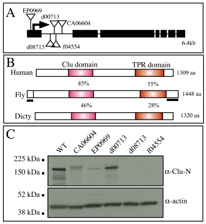
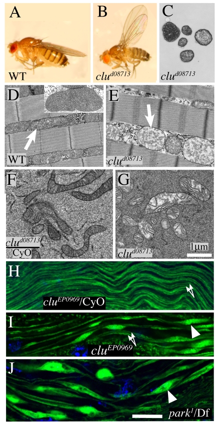
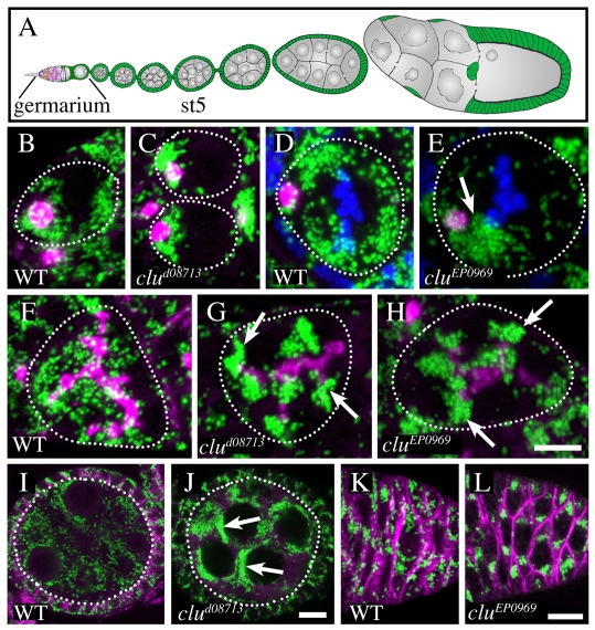
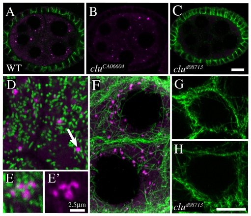
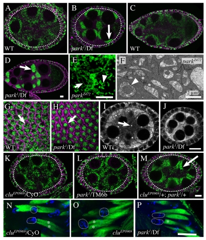
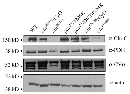
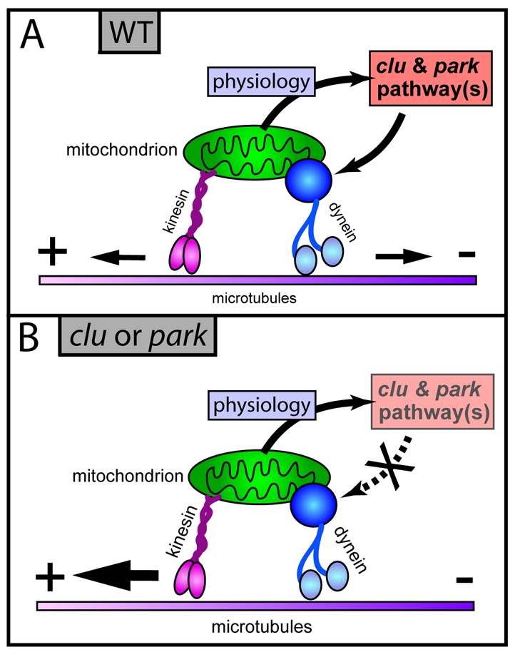
Parkinson's disease has been linked to altered mitochondrial function. Mutations in parkin (park), the Drosophila ortholog of a human gene that is responsible for many familial cases of Parkinson's disease, shorten life span, abolish fertility and disrupt mitochondrial structure. However, the role played by Park in mitochondrial function remains unclear. Here, we describe a novel Drosophila gene, clueless (clu), which encodes a highly conserved tetratricopeptide repeat protein that is related closely to the CluA protein of Dictyostelium, Clu1 of Saccharomyces cerevisiae and to similar proteins in diverse metazoan eukaryotes from Arabidopsis to humans. Like its orthologs, loss of Drosophila clu causes mitochondria to cluster within cells. We find that strong clu mutations resemble park mutations in their effects on mitochondrial function and that the two genes interact genetically. Conversely, mitochondria in park homozygotes become highly clustered. We propose that Clu functions in a novel pathway that positions mitochondria within the cell based on their physiological state. Disruption of the Clu pathway may enhance oxidative damage, alter gene expression, cause mitochondria to cluster at microtubule plus ends, and lead eventually to mitochondrial failure.


Figures

Fig. 1.
Fig. 1.
clu structure and expression. (A) Map of the clu locus showing sites of insertion mutation (triangles) and the translation start (arrow). Black boxes are exons. (B) Drosophila Clu compared with human Clu and Dictyostelium (Dicty) CluA. The position and percentage of amino acid identity of the Clu and TPR domains are shown. The segments used to prepare N-terminal- and C-terminal-specific antibodies are indicated by solid black bars. (C) Western blot of adult extracts probed with N-terminal anti-Clu antibody showing the effect of clu mutations (listed at the top of the lanes) on the 160 kD Clu protein.
Fig. 2.
Fig. 2.
Mitochondrial defects in clu mutants. (A) Wild-type adult female. (B) clud08713 adult female showing reduced size and abnormal wing position. (C) Light micrograph showing persistent larval fat body cells from the abdominal hemolymph of a 4-day-old clud08713 mutant female. (D) Electron micrograph of normal flight muscle showing mitochondria (arrow, inset). (E) clud08713 flight muscle showing abnormal, swollen mitochondria with extensive vacuolization of the inner membrane (arrow). (F) Normal mitochondria from clud08713/CyO ovarian germ cells. (G) Abnormal mitochondria from clud08713 ovaries showing partial vacuolization of the inner membrane. (H–J) Complex V-alpha subunit (CVα) expression (green) labeling mitochondrial derivatives in elongating spermatids from cluEP0969/CyO (H), cluEP0969 (I) and park1/Df (J) flies. In contrast to the uniform, paired mitochondrial derivatives that are seen normally (H, arrows), the mitochondria in both mutant animals (I,J) are uneven with swollen regions (arrowheads). Nuclei are stained with DAPI (blue). Bars, 1 μm (D–G); 5 μm (H–J).
Fig. 3.
Fig. 3.
clu mutations cause mitochondrial aggregation. (A) Diagram of a Drosophila ovariole, showing the nurse cells (gray), follicle cells (green) and progressively maturing follicles. The location of the germarium and a stage 5 (st5) follicle are indicated. In the germarium, germline stem cells (GSCs) contain a specialized organelle called the spectrosome that grows and interconnects the 16-cell germline cysts, where it is called the fusome (magenta). (B) Wild-type (WT) GSC showing dense mitochondria (green) at the anterior near the spectrosome (magenta circle), as well as throughout the cytoplasm. Note: the nucleus occupies the center of the cell. (C) clud08713 GSCs showing increased mitochondrial clustering around spectrosomes. (D,E) Mitochondria disperse across the spindle of a wild-type single germ cell (D), but remain clustered in a similar mitotic cluEP0969 germ cell (E, arrow). DAPI (blue) stains the metaphase plate. (F–H) Mitochondria disperse throughout the cytoplasm of early wild-type cysts (F), but cluster at the ends of the fusome in clud08713 (G) and cluEP0969 (H) mutant cysts. (I,J) Mitochondria are dispersed throughout the cytoplasm of stage 5 nurse cells in a wild-type cyst (I), but cluster (arrows) in clud08713 nurse cells (J). (K,L) Mitochondria in wild-type germarial follicle cells are dispersed (K), but clump extensively in cluEP0969 germarial follicle cells (L). White dashed line outlines indicate germ cells (B–E) or germline cysts (F–J). Fusome and lateral membranes are stained magenta (1B1); mitochondria are stained green (CVα). Bars, 10 μm.
Fig. 4.
Fig. 4.
Clu is a cytoplasmic, not mitochondrial, protein. (A–C) Stage 5 follicles: in wild-type follicles (A), Clu antibody (magenta) stains the nurse cell cytoplasm including some particles. Lateral membranes are stained green (1B1). The distribution of Clu-GFP in the protein trap line CA06604 (B), visualized with anti-GFP antibody (magenta), was indistinguishable from the distribution in wild-type follicles (A). In clud08713, Clu antibody staining is absent (C). (D–H) Stage 9 follicles: double label of Clu (magenta, arrow) and mitochondria (green) shows that Clu particles associate with one or more mitochondria (D). Juxtaposition of mitochondria (E, green) and Clu protein (E,E’, magenta) can be seen more clearly at higher magnification. (F) Association of microtubules (green) with some Clu bodies (magenta). (G,H) Microtubules in clud08713 stage 9 nurse cells (H) appear normal compared with a wild-type control (G). Bars, 10 μm (A–D,F–H); 2.5 μm (E’,E).
Fig. 5.
Fig. 5.
park mutations cause mitochondrial clustering and interact genetically with clu. (A) Wild-type stage 5 follicles lack clustered mitochondria (green). Lateral membranes are stained magenta (1B1). (B) Severe mitochondrial clustering in a stage 5 park1/Df follicle. (C) Wild-type stage 9 follicles lack clustered mitochondria. (D) Extreme mitochondrial clustering in a stage 9 park1/Df follicle. (E) The mitochondria in parkZ472 follicles are clustered, long (arrow) and frequently form ring structures (arrowhead). (F) Electron micrograph of a parkZ472 mitochondrial cluster showing doughnut-shaped mitochondria (arrowhead). (G) Wild-type stage 9 follicle cells mostly lack clustered mitochondria (arrow). (H) Mitochondria cluster in the cell center in most park1/Df stage 9 follicle cells (arrow). (I,J) Clu protein (white) is present at similar levels in wild-type (I) and park1/Df (J) follicles, but large Clu particles are absent in the mutant. (K–M) Normal dispersed mitochondrial distribution in cluEP0969/CyO (K) and park1/TM6b (L) stage 5 follicles. (M) Clustered mitochondria in a stage 5 follicle from a cluEP0969/+; park1/+ female. (N–P) Mitochondrial derivatives in cluEP0969/CyO (N), cluEP0969 (O) and park1/Df (P) male germ cells at the leaf blade stage. In wild-type spermatids (N), the Nebenkern unfurls revealing two mitochondria (asterisks) elongating below the nucleus (dotted circle). These mitochondria are less compact and appear frayed in cluEP0969 mutants (O). park1/Df mutants (P) have only one mitochondrion (asterisk) that looks smooth as in N. Mitochondria are stained green (CVα); membranes are stained magenta (1B1); nuclei are stained blue (DAPI). Bars, 10 μm, (A–D,G–M); 5 μm (E,N–P); 2 μm (F).
Fig. 6.
Fig. 6.
clu adults contain reduced amounts of mitochondrial protein. (A) Western blot of whole fly extracts probed for Clu, Pyruvate dehydrogenase (PDH), CVα and Actin. The level of the two mitochondrial proteins PDH and CVα were unchanged from wild-type levels in heterozygotes that were mutant for clu or park. However, both PDH and CVα levels were reduced significantly relative to the Actin control in strong clud08713 mutants (but not in cluEP0969).
Fig. 7.
Fig. 7.
Model for clu and park function. The figure shows a model for the role of the clu and park pathway(s) in cells (such as early ovarian germ cells) in which mitochondrial movement is controlled by the regulated engagement of plus-end-directed Kinesin motors (pink) and minus-end-directed Dynein motors (blue). The clu and park pathway(s) are located in the cytoplasm where they are proposed to sense the physiological state of a mitochondrion and modify its movement accordingly. (A) Under normal conditions, modulating the level of Dynein engagement relative to plus-end-directed motor activity allows the clu and park pathway(s) to cause a mitochondrion to move in either direction (arrows). (B) When the functioning of the clu and park pathway(s) is defective, or when levels of reactive oxygen species are elevated, minus end microtubule movement is blocked, causing mitochondria to move preferentially toward microtubule plus ends (large arrow). Note that in other cells, the clu and park pathway(s) might regulate mitochondrial movement by linking to different motors and transport systems.

Similar articles

Cited by

References

    1. Abou-Sleiman PM, Muqit MM, Wood NW. (2006). Expanding insights of mitochondrial dysfunction in Parkinson’s disease. Nat Rev Neurosci. 7, 207–219 - PubMed
    1. Allen AK, Spradling AC. (2008). The Sf1-related nuclear hormone receptor Hr39 regulates Drosophila female reproductive tract development and function. Development 135, 311–321 - PubMed
    1. Betarbet R, Sherer TB, Greenamyre JT. (2005). Ubiquitin-proteasome system and Parkinson’s diseases. Exp Neurol. 191, S17–S27 - PubMed
    1. Buszczak M, Spradling AC. (2006). The Drosophila P68 RNA helicase regulates transcriptional deactivation by promoting RNA release from chromatin. Genes Dev. 20, 977–989 - PMC - PubMed
    1. Buszczak M, Paterno S, Lighthouse D, Bachman J, Plank J, Owen S, Skora AD, Nystul TG, Ohlstein B, Allen A, et al. (2007). The Carnegie protein trap library: a versatile tool for Drosophila developmental studies. Genetics 175, 1505–1531 - PMC - PubMed

Publication types

MeSH terms