

Epstein-Barr virus infection is not a characteristic feature of multiple sclerosis brain
- PMID: 19638446
- PMCID: PMC2792367
- DOI: 10.1093/brain/awp200
Epstein-Barr virus infection is not a characteristic feature of multiple sclerosis brain
Abstract
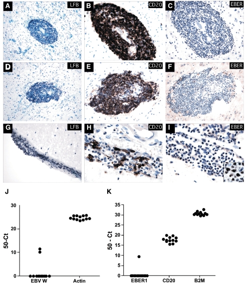
Multiple sclerosis is an inflammatory demyelinating disease of the central nervous system (CNS) that is thought to be caused by a combination of genetic and environmental factors. To date, considerable evidence has associated Epstein-Barr virus (EBV) infection with disease development. However, it remains controversial whether EBV infects multiple sclerosis brain and contributes directly to CNS immunopathology. To assess whether EBV infection is a characteristic feature of multiple sclerosis brain, a large cohort of multiple sclerosis specimens containing white matter lesions (nine adult and three paediatric cases) with a heterogeneous B cell infiltrate and a second cohort of multiple sclerosis specimens (12 cases) that included B cell infiltration within the meninges and parenchymal B cell aggregates, were examined for EBV infection using multiple methodologies including in situ hybridization, immunohistochemistry and two independent real-time polymerase chain reaction (PCR) methodologies that detect genomic EBV or the abundant EBV encoded RNA (EBER) 1, respectively. We report that EBV could not be detected in any of the multiple sclerosis specimens containing white matter lesions by any of the methods employed, yet EBV was readily detectable in multiple Epstein-Barr virus-positive control tissues including several CNS lymphomas. Furthermore, EBV was not detected in our second cohort of multiple sclerosis specimens by in situ hybridization. However, our real-time PCR methodologies, which were capable of detecting very few EBV infected cells, detected EBV at low levels in only 2 of the 12 multiple sclerosis meningeal specimens examined. Our finding that CNS EBV infection was rare in multiple sclerosis brain indicates that EBV infection is unlikely to contribute directly to multiple sclerosis brain pathology in the vast majority of cases.
Figures


Comment in
-
Epstein Barr virus is not a characteristic feature in the central nervous system in established multiple sclerosis.Brain. 2010 May;133(Pt 5):e137. doi: 10.1093/brain/awp296. Epub 2009 Nov 16. Brain. 2010. PMID: 19917644 No abstract available.
-
Detection of Epstein-Barr virus and B-cell follicles in the multiple sclerosis brain: what you find depends on how and where you look.Brain. 2010 Dec;133(Pt 12):e157. doi: 10.1093/brain/awq223. Epub 2010 Aug 25. Brain. 2010. PMID: 20739348 No abstract available.
References
-
- Alotaibi S, Kennedy J, Tellier R, Stephens D, Banwell B. Epstein-Barr virus in pediatric multiple sclerosis. JAMA. 2004;291:1875–9. - PubMed
-
- Alspaugh MA, Shoji H, Nonoyama M. A search for rheumatoid arthritis-associated nuclear antigen and Epstein-Barr virus specific antigens or genomes in tissues and cells from patients with rheumatoid arthritis. Arthritis Rheum. 1983;26:712–20. - PubMed
-
- Alvarez-Lafuente R, Garcia-Montojo M, De Las Heras V, Dominguez-Mozo M, Bartolome M, Benito-Martin M, et al. Herpesviruses and human endogenous retroviral sequences in the cerebrospinal fluid of multiple sclerosis patients. Mult Scler. 2008;14:595–601. - PubMed
-
- Anagnostopoulos I, Hummel M, Stein H. Frequent presence of latent Epstein-Barr virus infection in peripheral T cell lymphomas. A review. Leuk Lymphoma. 1995;19:1–12. - PubMed
-
- Ascherio A, Munger KL. Environmental risk factors for multiple sclerosis. Part I: the role of infection. Ann Neurol. 2007;61:288–99. - PubMed
Publication types
MeSH terms
Substances
Grants and funding
LinkOut - more resources
Full Text Sources
Medical
