. 2009 Jul;21(7):2036-44.
doi: 10.1105/tpc.109.068007. Epub 2009 Jul 28.

A structural basis for the pH-dependent xanthophyll cycle in Arabidopsis thaliana

Affiliations

A structural basis for the pH-dependent xanthophyll cycle in Arabidopsis thaliana

Pascal Arnoux et al. Plant Cell. 2009 Jul.

Abstract

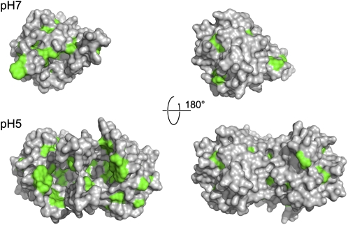
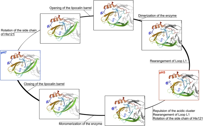
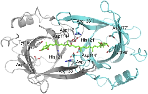
Plants adjust their photosynthetic activity to changing light conditions. A central regulation of photosynthesis depends on the xanthophyll cycle, in which the carotenoid violaxanthin is converted into zeaxanthin in strong light, thus activating the dissipation of the excess absorbed energy as heat and the scavenging of reactive oxygen species. Violaxanthin deepoxidase (VDE), the enzyme responsible for zeaxanthin synthesis, is activated by the acidification of the thylakoid lumen when photosynthetic electron transport exceeds the capacity of assimilatory reactions: at neutral pH, VDE is a soluble and inactive enzyme, whereas at acidic pH, it attaches to the thylakoid membrane where it binds its violaxanthin substrate. VDE also uses ascorbate as a cosubstrate with a pH-dependent Km that may reflect a preference for ascorbic acid. We determined the structures of the central lipocalin domain of VDE (VDEcd) at acidic and neutral pH. At neutral pH, VDEcd is monomeric with its active site occluded within a lipocalin barrel. Upon acidification, the barrel opens up and the enzyme appears as a dimer. A channel linking the two active sites of the dimer can harbor the entire carotenoid substrate and thus may permit the parallel deepoxidation of the two violaxanthin beta-ionone rings, making VDE an elegant example of the adaptation of an asymmetric enzyme to its symmetric substrate.


Figures

Figure 1.
Figure 1.
Sequence Analysis of VDE. (A) Schematic domain organization of VDE. (B) Sequence alignment of a range of VDE enzymes. Conserved residues are color coded according to the domain organization. Secondary structure features are shown and labeled above the alignment (cylinders indicate α-helices and arrows β-sheets); residues important for the pH switch are marked with black stars and residues forming the putative active site with black squares. Residues at the dimer interface are boxed.
Figure 2.
Figure 2.
Architecture of the Central Lipocalin Domain of VDE at pH 7 and 5. Crystal structure of VDEcd at pH 7.0 (A) and pH 5.0 (B) with amino acid sequence variation mapped onto the surface view (right). The opening of the top and side of the barrel at pH 5.0 is visible in this surface view. Membrane anchoring is expected to occur on this side of the molecule due to the abundance of hydrophobic residues (see also Figure 5).
Figure 3.
Figure 3.
pH-Dependent Dimerization and Opening of the VDE Active Sites. Positions of key conserved residues at pH 7.0 (A) and pH 5.0 (B) are shown. Note the rotation of the side chain of His-121 and the rearrangement of the L1 loop with Asp-114 hydrogen bonded to Tyr-198 at neutral pH, whereas it is clustered with Asp-117, Asp-114, and Asp-117 from the adjoining monomer at acidic pH.
Figure 4.
Figure 4.
Structural Comparison of the Open and Closed Forms of VDEcd. (A) Superposition of the VDEcd structure obtained at pH 7.0 (green) with the VDEcd structure obtained at pH 5.0 (one monomer colored in blue and the other colored in gray and rendered semitransparent). (B) Root mean square deviation plot between the structures obtained at pH 7 and 5. Secondary structures and their labeling are depicted above the plot.
Figure 5.
Figure 5.
Position of Conserved Hydrophobic Residues at Neutral (Top) and Acidic pH (Bottom). Surface representation of VDEcd with conserved hydrophobic residues (Ala, Val, Ile, Leu, Phe, Tyr, and Trp) colored in green. Being more exposed to the solvent at acidic pH may play a role in the attachment of VDE to the membrane.
Figure 6.
Figure 6.
Plausible Transitions between the Closed (pH 7) and Open (pH 5) States of VDEcd. Residues discussed in the text as being important for the pH transition are represented in stick form. The adjoining monomer is shown in gray and is either transparent or solid depending on the estimated plausibility of the structure. Pauses separate His-121 reorientation, barrel opening, loop rearrangements, and dimer formation. For the neutral to acidic pH transition, the His-121 side chain rotation and barrel opening are probably the first events. Transition in the reverse order (rearrangement of loop L1 followed by dimer formation and strand opening) would be disallowed, as the closed barrel structure would prevent dimer formation. For the acidic-to-neutral pH transition, repulsion within the acidic clusters (Asp-114 and Asp-117 from both monomers) and rearrangement of loop1 are the most likely primary events, as otherwise steric clashes would prevent the barrel from closing before loop1 rearranges.
Figure 7.
Figure 7.
Enzymatic Activity of VDE Mutants. The enzymatic activity of VDE mutants is indicated as a percentage of that of the wild type and quantified from an increase in absorption at 502 nm (Yamamoto, 1985; Bugos et al., 1999). Data are expressed as the means ± sd of six independent experiments. In the case of inactive mutants, the absence of Z was confirmed by HPLC. Protein concentration was estimated by protein gel blot analysis before testing their activity. [See online article for color version of this figure.]
Figure 8.
Figure 8.
Model of V Docking on the VDEcd Dimer. Approximate model of the interaction between VDEcd and V. The V structure was taken from the x-ray structure of the spinach light-harvesting complex LHCII (Liu et al., 2004) without modification and manually docked into the dimeric structure obtained at pH 5. The ascorbate cosubstrate was not modeled, but there is enough space in the central lipocalin cavity for this cosubstrate, even in the presence of V.

References

    1. Ahn, T.K., Avenson, T.J., Ballottari, M., Cheng, Y.C., Niyogi, K.K., Bassi, R., and Fleming, G.R. (2008). Architecture of a charge-transfer state regulating light harvesting in a plant antenna protein. Science 320 794–797. - PubMed
    1. Amarie, S., Laura, W., Tiago, B., Werner, K., Andreas, D., and Josef, W. (2009). Properties of zeaxanthin and its radical cation bound to the minor light-harvesting complexes CP24, CP26 and CP29. Biochimic. Biophys. Acta 1787 747–752. - PubMed
    1. Barber, J., and Andersson, B. (1992). Too much of a good thing: Light can be bad for photosynthesis. Trends Biochem. Sci. 17 61–66. - PubMed
    1. Bratt, C., Arvidsson, P., Carlsson, M., and Akerlund, H. (1995). Regulation of violaxanthin de-epoxidase activity by pH and ascorbate. Photosynth. Res. 45 169–175. - PubMed
    1. Bugos, R.C., Chang, S.H., and Yamamoto, H.Y. (1999). Developmental expression of violaxanthin de-epoxidase in leaves of tobacco growing under high and low light. Plant Physiol. 121 207–214. - PMC - PubMed

MeSH terms