. 2009 Oct;76(4):824-32.
doi: 10.1124/mol.109.058883. Epub 2009 Jul 28.

An allosteric mechanism for inhibiting HIV-1 integrase with a small molecule

Affiliations

An allosteric mechanism for inhibiting HIV-1 integrase with a small molecule

Jacques J Kessl et al. Mol Pharmacol. 2009 Oct.

Abstract

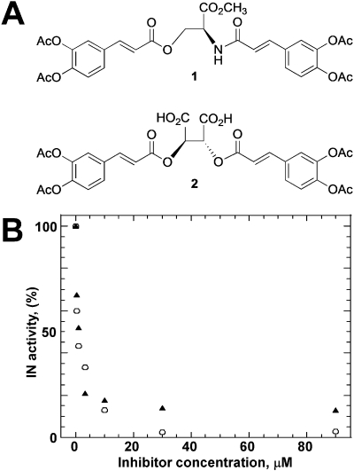
HIV-1 integrase (IN) is a validated target for developing antiretroviral inhibitors. Using affinity acetylation and mass spectrometric (MS) analysis, we previously identified a tetra-acetylated inhibitor (2E)-3-[3,4-bis(acetoxy)phenyl]-2-propenoate-N-[(2E)-3-[3,4-bis(acetyloxy)phenyl]-1-oxo-2-propenyl]-L-serine methyl ester; compound 1] that selectively modified Lys173 at the IN dimer interface. Here we extend our efforts to dissect the mechanism of inhibition and structural features that are important for the selective binding of compound 1. Using a subunit exchange assay, we found that the inhibitor strongly modulates dynamic interactions between IN subunits. Restricting such interactions does not directly interfere with IN binding to DNA substrates or cellular cofactor lens epithelium-derived growth factor, but it compromises the formation of the fully functional nucleoprotein complex. Studies comparing compound 1 with a structurally related IN inhibitor, the tetra-acetylated-chicoric acid derivative (2R,3R)-2,3-bis[[(2E)-3-[3,4-bis(acetyloxy)phenyl]-1-oxo-2-propen-1-yl]oxy]-butanedioic acid (compound 2), indicated striking mechanistic differences between these agents. The structures of the two inhibitors differ only in their central linker regions, with compounds 1 and 2 containing a single methyl ester group and two carboxylic acids, respectively. MS experiments highlighted the importance of these structural differences for selective binding of compound 1 to the IN dimer interface. Moreover, molecular modeling of compound 1 complexed to IN identified a potential inhibitor binding cavity and provided structural clues regarding a possible role of the central methyl ester group in establishing an extensive hydrogen bonding network with both interacting subunits. The proposed mechanism of action and binding site for the small-molecule inhibitor identified in the present study provide an attractive venue for developing allosteric inhibitors of HIV-1 IN.


Figures

Fig. 1.
Fig. 1.
A, structures of compounds 1 and 2. B, the inhibition profiles of recombinant wild-type IN with compounds 1 (▲) and 2 (○). The mean values for at least three experiments are presented. The deviation for each measurement was ±10%.
Fig. 2.
Fig. 2.
Segments of quadrupole/time of flight mass spectra demonstrating selective acetylation of Lys173 with 1. A, IN + 6.25 μM 1; B, IN + 25 μM 1; C, IN + 10 μM 2; D, IN + 200 μM 2; E, free IN. The tryptic peptide of IN containing acetylated Lys173 is indicated. C1 is unmodified tryptic peptide (AMASDFNLPPVVAK) of IN, which serves as an internal control.
Fig. 3.
Fig. 3.
Effects of 1 and 2 on IN-DNA cross-linking. IN(E152C) was first incubated with increasing concentrations of 1 (A) and 2 (B) and then cross-linked to a specific DNA containing a cross-linkable analog at the G2 position (G2). The reaction products were resolved by SDS-PAGE. The bands corresponding to free IN and IN-DNA complex are indicated. A, lane 1 molecular mass markers; lane 2, IN-DNA cross-linking in the absence of 1. Lanes 3 to 10 contained increasing concentrations of 1 (1, 2, 4, 8, 16, 32, 64, and 128 μM). B, lane 1, molecular mass markers; lane 2, IN-DNA cross-linking in the absence of 2; lanes 3 to 9 contained increasing concentrations of 2 (2, 4, 8, 16, 32, 64, and 128 μM).
Fig. 4.
Fig. 4.
Effects of 1 and 2 on IN subunit-subunit interactions. Experimental design (A) and results (B). A, the subunit exchange between IN multimers was tested by mixing the two wild-type IN proteins: IN1-IN1, a tag-free form, and IN2-IN2, containing the histidine tag at its C terminus. The full-length proteins are depicted as dimers (IN1–IN1) and (IN2–IN2). Upon subunit-subunit exchange, three IN populations can be formed: IN1–IN1, IN1–IN2, and IN2–IN2. Of these, IN2–IN2 and IN1–IN2 can be pulled down by nickel-affinity resin through binding with the histidine tag, whereas the tag-free IN1–IN1 is washed out. B, the IN1 and IN2 proteins from the bound complexes were subjected to SDS-PAGE and detected by Western blot. Lane 1, IN2 load; lane 2, IN1 load; lane 3, no ligand or IN1 (non specific pull-down control); lane 4, the subunit-subunit exchange in the absence of compounds; lanes 5 to 10, the subunit exchange reactions in the presence of increasing 1 concentrations (8, 16, 32, 64, 128, and 256 μM); lanes 11 and 12, reactions in the presence of 128 and 256 μM 2. C, order-of-addition experiments. Lanes 1 and 3, subunit exchange reactions in the absence of compounds; lane 2, IN2-IN2 was first preincubated with 1 and then exposed to IN1-IN1; lane 4, IN2-IN2 and IN1-IN1 were first mixed to carry out subunit exchange, and then 1 was added to the mixture.
Fig. 5.
Fig. 5.
Effects of 1 on IN multimerization (A), solubility (B), and LEDGF-IN binding (C). A, in parallel reactions, free IN (lane 1) and the IN-1 complex (lane 2) were subjected to cross-linking with BS3, and the reaction products were separated by SDS-PAGE. Migrations of molecular mass markers and IN bands are indicated. B, lane 1, molecular mass markers; lane 2, total (T) sample before centrifugation; lanes 3 and 4, supernatant (S) and precipitate (P) fractions formed after centrifugation of IN in the presence of 10% dimethyl sulfoxide; lanes 5 and 6, IN solubility in the presence of 256 μM 1. C, lane 1, LEDGF input; lane 2, no ligand or IN (nonspecific pull down control); lane 3, LEDGF and IN interactions in the absence of 1; lanes 4 to 8, assays with increasing 1 concentrations (8, 16, 32, 64, and 128 μM).
Fig. 6.
Fig. 6.
Molecular docking studies for 1. A and B, two potential binding pockets (colored in magenta and orange) at the CCD dimer interface. These distinct sites are located immediately adjacent to one another. A and B, two different views of the CCD dimer to better visualize individual binding pockets. Individual subunits of IN are colored green and yellow. The location of Lys173 and height (H), depth (D), and width (W) for each cavity is indicated. C, the space-filling model for the CCD dimer-1 complex showing that the inhibitor simultaneously interacts with both IN monomers (green and yellow). The central methyl ester group is deeply buried in the cavity and cannot be seen in this picture. Integrase active site residues (Asp64, Asp116, and Glu152) on each monomer are shown in red. D, key interactions with IN residues established by the central methyl ester group and the linker region. The amino acids from green or yellow subunits are colored accordingly.
Fig. 7.
Fig. 7.
Biochemical analysis of IN mutants. A, size-exclusion chromatography of wild-type and mutant proteins. Peaks corresponding to tetramer (Tet) IN with estimated molecular masses of ∼111 kDa and a dimeric (Dim) protein with estimate molecular masses of ∼54 kDa, are indicated. B, effects of amino acid substitutions on recombinant IN activities. Top image depicts strand transfer activities. Positions of 21-oligomer substrate (21-S) and reaction products (STP) are indicated. Bottom image displays 3′-processing activities. The positions of 21-S and specific 19-oligomer products (19-P) are shown. B, LEDGF binding to wild-type and mutant INs: lane 1, molecular mass markers; lane 2, LEDGF input; lane 3, assay without IN (nonspecific pull-down control); lane 4, assay with wild-type IN; lane 5, assay with E87A mutant; lane 6, assay with E96A mutant; lane 7, assay with Y99A mutant; lane 8, assay with K103A mutant.

Similar articles

Cited by

References

    1. Al-Mawsawi LQ, Christ F, Dayam R, Debyser Z, Neamati N. (2008) Inhibitory profile of a LEDGF/p75 peptide against HIV-1 integrase: insight into integrase-DNA complex formation and catalysis. FEBS Lett 582:1425–1430 - PubMed
    1. Al-Mawsawi LQ, Fikkert V, Dayam R, Witvrouw M, Burke TR, Jr, Borchers CH, Neamati N. (2006) Discovery of a small-molecule HIV-1 integrase inhibitor-binding site. Proc Natl Acad Sci U S A 103:10080–10085 - PMC - PubMed
    1. Busschots K, De Rijck J, Christ F, Debyser Z. (2009) In search of small molecules blocking interactions between HIV proteins and intracellular cofactors. Mol Biosyst 5:21–31 - PubMed
    1. Chen JC, Krucinski J, Miercke LJ, Finer-Moore JS, Tang AH, Leavitt AD, Stroud RM. (2000) Crystal structure of the HIV-1 integrase catalytic core and C-terminal domains: a model for viral DNA binding. Proc Natl Acad Sci U S A 97:8233–8238 - PMC - PubMed
    1. Cherepanov P, Ambrosio AL, Rahman S, Ellenberger T, Engelman A. (2005) Structural basis for the recognition between HIV-1 integrase and transcriptional coactivator p75. Proc Natl Acad Sci U S A 102:17308–17313 - PMC - PubMed

Publication types

MeSH terms

Substances

LinkOut - more resources