. 2009 Aug 1;69(15):6282-9.
doi: 10.1158/0008-5472.CAN-09-1176. Epub 2009 Jul 28.

Inhibition of superoxide generation upon T-cell receptor engagement rescues Mart-1(27-35)-reactive T cells from activation-induced cell death

Affiliations

Inhibition of superoxide generation upon T-cell receptor engagement rescues Mart-1(27-35)-reactive T cells from activation-induced cell death

Håkan Norell et al. Cancer Res. .

Abstract

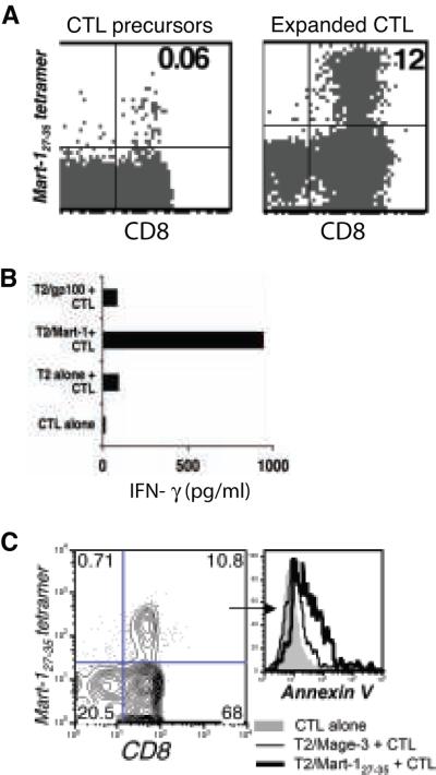
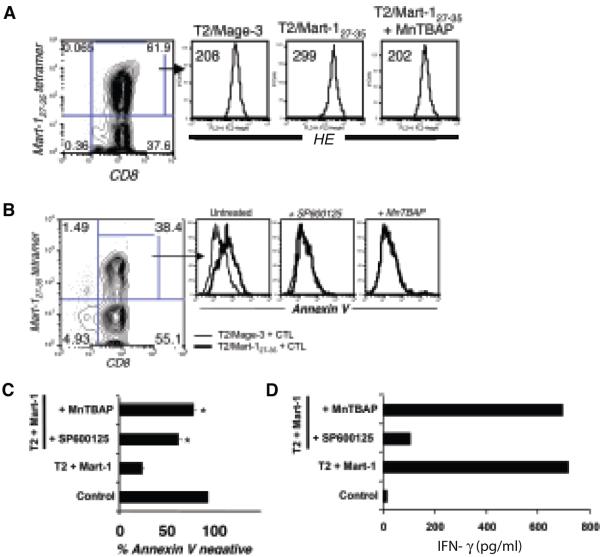
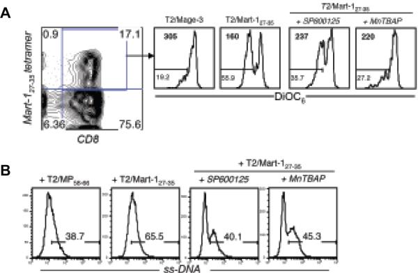
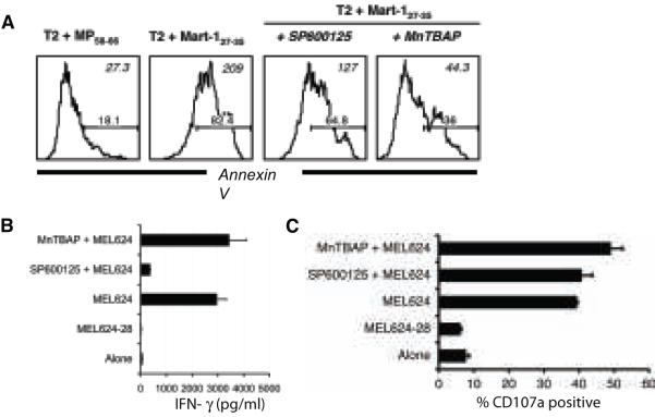
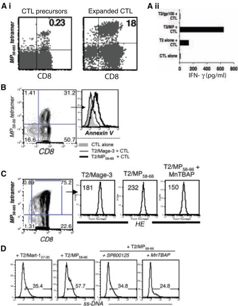
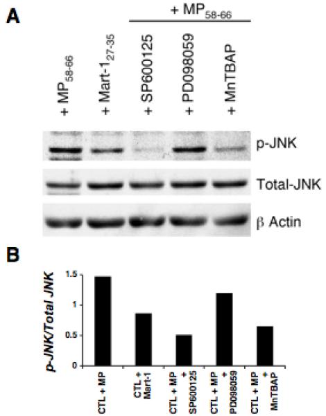
Cytotoxic T lymphocytes (CTL) may undergo massive expansion upon appropriate antigenic stimulation. Homeostasis is maintained by a subsequent "contraction" of these cells. Activation-induced cell death (AICD) and programmed cell death prevent the untoward side effects, arising from excessive numbers and prolonged persistence of activated CTL, that occur upon uncontrolled and/or continued expansion. However, effector cell persistence has been identified as a hallmark of successful T-cell-mediated adoptive immunotherapy. Thus, prevention of AICD may be critical to achieve more successful clinical results. We have previously shown that treatment with the c-Jun NH(2)-terminal kinase (JNK) inhibitor SP600125 protects human melanoma epitope Mart-1(27-35)-reactive CTL from apoptotic death upon their reencounter with cognate antigen. However, inhibition of JNK also interferes with the functional ability of the CTL to secrete IFN-gamma. Here, we show that reactive oxygen species (ROS) inhibitors, such as the superoxide dismutase mimetic Mn (III) tetrakis (5, 10, 15, 20-benzoic acid) porphyrin (MnTBAP), efficiently protected Mart-1(27-35)-reactive primary CTL from AICD without impairing their functional capability. MnTBAP prevented the increase in intracellular ROS, mitochondrial membrane collapse, and DNA fragmentation observed in control-treated cells upon cognate antigen encounter. Furthermore, the mechanism of AICD prevention in primary CTL included blockade of JNK activation. Finally, tumor-reactive in vitro expanded tumor infiltrating lymphocytes, which are used clinically in cancer immunotherapy, also benefit from MnTBAP-mediated antioxidant treatment. Thus, modulation of the redox pathway might improve CTL persistence and lead to better clinical results for T cell-based immunotherapies.


Figures

Figure 1
Figure 1. Expansion of Mart-127-35 reactive CTL and induction of AICD
(A) Mart-127-35 CTL precursors (left) and CTL expanded with autologous DC pulsed with peptides were tracked by FACS using staining for CD8 and tetramer reagents. (B) IFN-γ secreted into co-culture supernatant collected after overnight incubation of Mart-127-35 CTL stimulated with cognate or control peptide-pulsed T2 cells. (C) Expanded CTL were restimulated with cognate or control epitope-pulsed T2 cells and were stained after 4 hours with tetramer, CD8, and Annexin V. Histogram depict fluorescence intensity for Annexin V in tetramer-gated CD8+ CTL when unstimulated (grey filled), stimulated with T2 cells pulsed with control peptide (thin black), and stimulated with T2 cells pulsed with cognate peptide (thick black). All data represents one of at least seven separate experiments with similar results.
Figure 2
Figure 2. Increased endogenous ROS levels after TCR stimulation and rescue of Mart-127-35 reactive CTL from AICD by ROS inhibition
Mart-127-35 epitope-reactive CTL were stimulated with control or cognate peptide-pulsed T2 cells (1 μg/ml) or first preincubated with 400 μM of MnTBAP or 25 μM SP600125 for 30 min and then stimulated with their cognate epitope. (A). Staining with hydro ethidium (HE) was performed 4 hours later. Histogram represents fluorescence intensity of HE on the antigen-reactive CTL. Numbers represent mean fluorescence intensity (MFI). (B). Induction of AICD in CTL was evaluated by staining for tetramer, CD8, and Annexin V 4 hours later and analysis using FACS. Histogram overlays depict Annexin V expression on tetramer-gated CTL stimulated with T2 cells pulsed with control peptide (thin black) and relevant peptide (thick black), respectively. MAGE-3 (control epitope); Mart-127-35 (cognate epitope). (C) Another representation of how Mart-127-35 reactive CTL were rescued from death (and thus remained Annexin V negative) to various degrees in response to pretreatment with SP600125 and MnTBAP before cognate antigen exposure. * p<0.05, as compared to untreated CTL stimulated with T2 cells pulsed with Mart-1 peptide. (D) Matched IFN-γ secretion from the setup in (C) measured by ELISA. All data represent one of three separate experiments with similar results.
Figure 3
Figure 3. Effect of TCR stimulation and ROS inhibition on the membrane potential (Δψ) and DNA degradation
Mart-127-35 reactive CTL preincubated with MnTBAP (400 μM) and SP600125 (25 μM) were stimulated with cognate Mart-127-35 and control Mage-3 peptide-pulsed T2 cells for induction of AICD. (A) Histogram represents DiOC6 staining on tetramer-gated CTL. Numbers in the upper left corner represent MFI for DiOC6 staining and in the lower left corner represent the percentage of cells that have low membrane potential. (B) ss-DNA levels in tetramer-positive cells were determined with ss-DNA-specific antibody. Data presented show one representative experiments of two.
Figure 4
Figure 4. Pretreatment with MnTBAP protects TIL from AICD without impairing function
Mart-1- reactive TIL1235 preincubated with media alone, MnTBAP (250-500 μM) and SP600125 (25 μM) were co-cultured with target cells. (A) After 4 hours co-culture with T2 cells pulsed with cognate or control peptide (1 μg/ml) cells were stained with Annexin V, 7AAD and CD8 and analyzed by FACS. Histogram represent the log fluorescence of Annexin V on CD8+ gated TIL. Numbers in the upper right corner represent MFI and in the lower right corner represent the percentage of cells that stained positive for Annexin V. (B) IFN-γ release by and (C) CD107a expression on TIL1235 upon co-culture with HLA-A2-positive (MEL624) or matched HLA-A2-negative (MEL624-28) melanoma cells. All data shown are from one representative experiment of at least two.
Figure 5
Figure 5. Effect of MnTBAP on AICD of influenza epitope MP58-66 reactive T cells
(Ai). Tetramer staining to track CTL precursors (left) that were expanded using MP58-66 peptide-pulsed DC and stained on day 12 (right). (Aii) IFN-γ secretion after overnight incubation of the MP58-66 CTL stimulated with cognate or control peptide gp100209 pulsed T2 cells. Data shown in (A) represent one of more than five separate experiments with similar results. (B) Histogram overlays depict fluorescence intensity for Annexin V in MP58-66 tetramer-gated CTL when unstimulated (grey filled), stimulated using T2 cells pulsed with control Mage-3 peptide (thin black), and stimulated using T2 cells pulsed with cognate MP58-66 peptide (thick black). (C) MP58-66 -reactive CTL untreated or pretreated with MnTBAP were stimulated with control Mage-3 or cognate MP58-66 peptide-pulsed (1 μg/ml) T2 cells. Staining was performed 4 hours later. Histogram represents fluorescence intensity of HE on the antigen-reactive CTL. Numbers in upper left corner represent MFI. Data in (B) and (C) represent one of three separate experiments with similar results. (D). Untreated, MnTBAP and SP600125 pretreated MP58-66 epitope-reactive CTL were exposed to the cognate (MP58-66) and control (Mart-127-35) peptide-pulsed T2 cells for induction of AICD. Histogram represents ss-DNA levels in tetramer-positive cells determined after staining with ss-DNA specific antibody. Data from one representative experiment of two is shown.
Figure 6
Figure 6. Effect of ROS inhibition on JNK phosphorylation
MP58-66 reactive SP600125, MnTBAP or PD098059 (ERK inhibitor) pretreated CTL were exposed to the cognate epitope MP58-66 and untreated CTL were exposed to both cognate epitope MP58-66 and control epitope Mart-127-35 for induction of AICD. (A) Western blot depicts total and phosphorylated (p-)JNK levels using lysates made from the MP58-66 reactive CTL. (B) Densitometric analysis of the blot in (A). Bands were scanned into the computer and band intensity was quantitated using the National Institutes of Health (NIH) Image 1.61 software (free software available from the NIH). Values represent the ratio of p-JNK over total JNK.

Comment in

  • Immunotherapy. 2009 Nov;1(6):921-4

Similar articles

Cited by

References

    1. Jaattela M. Programmed cell death: many ways for cells to die decently. Ann Med. 2002;34:480–8. - PubMed
    1. Hildeman D, Jorgensen T, Kappler J, Marrack P. Apoptosis and the homeostatic control of immune responses. Curr Opin Immunol. 2007;19:516–21. - PMC - PubMed
    1. Lu B, Finn OJ. T-cell death and cancer immune tolerance. Cell Death Differ. 2008;15:70–9. - PubMed
    1. Whiteside TL. Apoptosis of immune cells in the tumor microenvironment and peripheral circulation of patients with cancer: implications for immunotherapy. Vaccine. 2002;20(Suppl 4):A46–51. - PubMed
    1. Huang J, Khong HT, Dudley ME, et al. Survival, persistence, and progressive differentiation of adoptively transferred tumor-reactive T cells associated with tumor regression. J Immunother. 2005;28:258–67. - PMC - PubMed

Publication types

MeSH terms