. 2009 Oct 1;114(14):3033-43.
doi: 10.1182/blood-2009-02-204966. Epub 2009 Jul 28.

Syndecan-1 shedding facilitates the resolution of neutrophilic inflammation by removing sequestered CXC chemokines

Affiliations

Syndecan-1 shedding facilitates the resolution of neutrophilic inflammation by removing sequestered CXC chemokines

Kazutaka Hayashida et al. Blood. .

Abstract

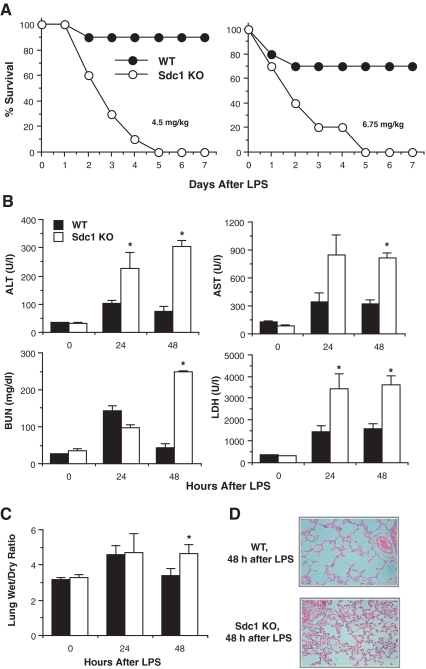
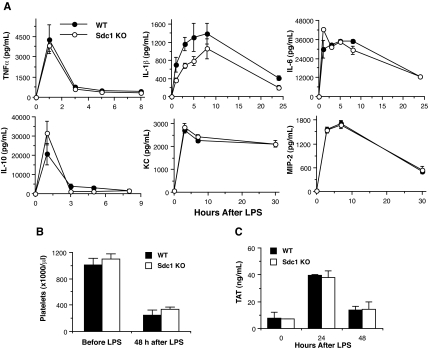
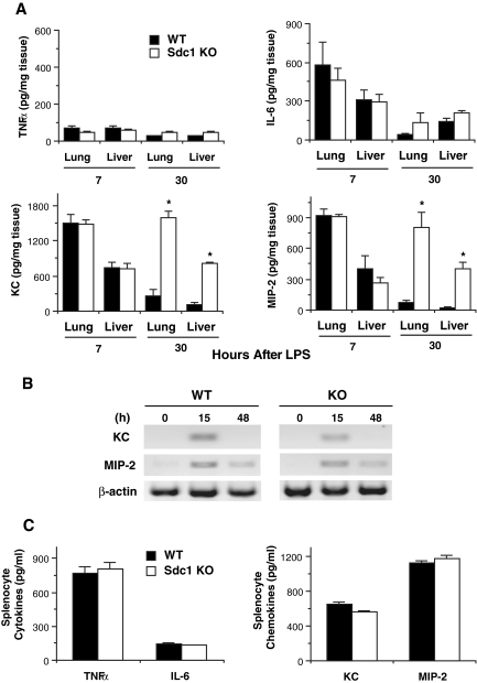
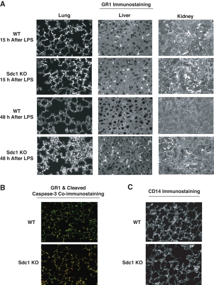
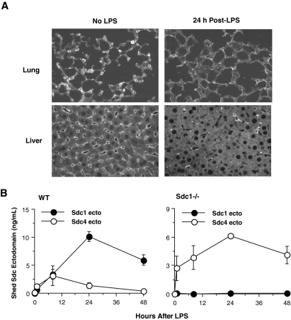
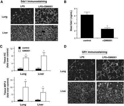
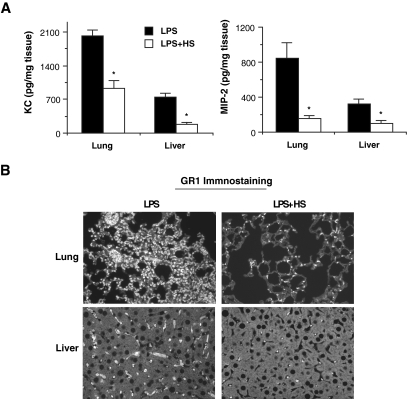
Heparan sulfate binds to and regulates many inflammatory mediators in vitro, suggesting that it serves an important role in directing the progression and outcome of inflammatory responses in vivo. Here, we evaluated the role of syndecan-1, a major heparan sulfate proteoglycan, in modulating multiorgan host injury responses in murine endotoxemia. The extent of systemic inflammation was similar between endotoxemic syndecan-1-null and wild-type mice. However, high levels of CXC chemokines (KC and MIP-2), particularly at later times after LPS, were specifically sustained in multiple organs in syndecan-1-null mice and associated with exaggerated neutrophilic inflammation, organ damage, and lethality. Syndecan-1 shedding was activated in several organs of endotoxemic wild-type mice, and this associated closely with the removal of tissue-bound CXC chemokines and resolution of accumulated neutrophils. Moreover, administration of a shedding inhibitor exacerbated disease by impeding the removal of CXC chemokines and neutrophils, whereas administration of heparan sulfate inhibited the accumulation of CXC chemokines and neutrophils in tissues and attenuated multiorgan injury and lethality. These data show that syndecan-1 shedding is a critical endogenous mechanism that facilitates the resolution of neutrophilic inflammation by aiding the clearance of proinflammatory chemokines in a heparan sulfate-dependent manner.


Figures

Figure 1
Figure 1
Sdc1−/− mice are susceptible to LPS-induced multiorgan injury and dysfunction and lethal endotoxemia. (A) WT and Sdc1−/− mice were injected intraperitoneally with 4.5 or 6.75 mg LPS/kg, and their survival was tracked for 7 days (n = 10 in each group; P < .05 at ≥ 3 days after LPS at both LPS doses). At a higher dose of LPS (15 mg/kg), all WT and Sdc1−/− mice died by 3 days after LPS, whereas at a lower dose (2.25 mg/kg), all WT and Sdc1−/− mice survived (not shown). (B) WT and Sdc1−/− mice were injected with LPS (4.5 mg/kg) and serum levels of ALT, AST, BUN, and LDH were measured at 0, 24, and 48 hours after LPS by a serum chemistry analyzer (Cobas Integra 400 Plus serum chemistry analyzer). Results shown are mean ± SE (n = 5; *P < .05 relative to WT mice). (C) WT and Sdc1−/− mice were injected with LPS, and the lung wet/dry ratio was determined at the indicated times by weighing lungs before and after incubation at 90°C for 3 days (n = 5). (D) Paraffin-embedded tissues sections (5 μm) of WT and Sdc1−/− lungs harvested at 48 hours after LPS were stained with hematoxylin-eosin (original magnification, ×200). Error bars indicate SE.
Figure 2
Figure 2
Endotoxemic WT and Sdc1−/− mice show similar systemic inflammatory and hemostatic responses. Mice were injected with LPS, and (A) serum levels of TNFα, IL-1β, IL-6, IL-10, KC, and MIP-2 were determined by ELISA at the indicated times after LPS (n = 5), (B) circulating platelets were measured by CBC analysis before and 48 hours after LPS (n = 3; Bayer Advia 120 hematology analyzer), and (C) plasma TAT levels were determined by ELISA at the indicated times after LPS (n = 4). Error bars indicate SE.
Figure 3
Figure 3
Removal of tissue-associated KC and MIP-2 is impeded in endotoxemic Sdc1−/− mice. (A) WT and Sdc1−/− mice were injected with LPS, and their lungs and livers were harvested at 7 or 30 hours after LPS. Tissues were weighed and homogenized, and tissue levels of TNFα, IL-6, KC, and MIP-2 were determined by ELISA (n = 5; *P < .05 relative to WT mice at the indicated time). (B) Total RNA was isolated from WT and Sdc1−/− lungs at 0, 15, and 48 hours after LPS infusion, and KC, MIP-2, and β-actin mRNA was assessed by reverse transcription polymerase chain reaction. (C) WT or Sdc1−/− splenocytes were stimulated with 100 ng LPS/mL for 24 hours at 37°C, and the concentration of TNFα, IL-6, KC, and MIP-2 in the conditioned medium was determined by ELISA (n = 4). Error bars indicate SE.
Figure 4
Figure 4
Resolution of neutrophilic inflammation is inhibited in endotoxemic Sdc1−/− mice. (A) WT and Sdc1−/− mice were injected with LPS, and their lungs, livers, and kidneys were isolated at 15 or 48 hours after LPS. Tissue sections were immunostained with the rat anti–mouse GR-1 (Ly6G/C, clone RBC6-8C5) antibody and Alexa 594 donkey anti–rat IgG antibody (original magnification, ×200). Similar results were obtained with the rat anti–mouse Ly6G (1A8) monoclonal antibody (not shown). (B) WT and Sdc1−/− lung sections (24 hours after LPS) were coimmunostained with rabbit anti–cleaved caspase-3 and anti-GR1 antibodies and with Alexa 594 anti–rat and Alexa 488 goat anti–rabbit antibodies (original magnification, ×200). (C) WT and Sdc1−/− lung sections (24 hours after LPS) were immunostained with rat anti-CD14 and Alexa 594 anti–rat antibodies (original magnification, ×200).
Figure 5
Figure 5
Syndecan-1 ectodomains are shed from the cell surface in various organs and released into the circulation in endotoxemic WT mice. (A) WT mice were injected with or without LPS, and their lungs and livers were isolated at 24 hours after LPS. Tissue sections were immunostained with 281-2 rat anti–mouse syndecan-1 ectodomain and Alexa 594 donkey anti–rat IgG antibodies (original magnification, ×200). (B) WT and Sdc1−/− mice were injected with LPS, and serum levels of syndecan-1 and -4 ectodomains were determined at 0, 1, 8, 24, and 48 hours after LPS by dot immunoblotting (n = 5 at each time point). Error bars indicate SE.
Figure 6
Figure 6
Delayed GM6001 infusion inhibits syndecan-1 shedding, removal of CXC chemokines in tissues, and resolution of neutrophilic inflammation. WT mice were injected intraperitoneally with or without 150 mg GM6001/kg at 24 hours after LPS, and tissue samples were analyzed at 40 hours after LPS. Syndecan-1 shedding was assessed by (A) immunostaining lung and liver sections (original magnification, ×200) and (B) measuring serum levels of syndecan-1 ectodomains (n = 4). (C) Tissue levels of KC and MIP-2 were determined by ELISA (n = 4). (D) Neutrophil accumulation was assessed by immunostaining with anti-GR1 and Alexa 594 anti–rat antibodies (original magnification, ×200). Error bars indicate SE.
Figure 7
Figure 7
Delayed HS administration facilitates the removal of tissue-associated CXC chemokines and resolution of neutrophilic inflammation. (A) Sdc1−/− mice were injected intraperitoneally with or without 50 μg HS/mouse at 10 hours after LPS, and tissue samples were analyzed at 40 hours after LPS. Tissues levels of KC and MIP-2 were determined by ELISA (n = 5). (B) Neutrophil accumulation was assessed by immunostaining with anti-GR1 and Alexa 594 anti–rat antibodies (original magnification, ×200). Error bars indicate SE.

References

    1. Bernfield M, Götte M, Park PW, et al. Functions of cell surface heparan sulfate proteoglycans. Annu Rev Biochem. 1999;68:729–777. - PubMed
    1. Esko JD, Selleck SB. Order out of chaos: assembly of ligand binding sites in heparan sulfate. Annu Rev Biochem. 2002;71:435–471. - PubMed
    1. Tyrrell DJ, Horne AP, Holme KR, Preuss JM, Page CP. Heparin in inflammation: potential therapeutic applications beyond anticoagulation. Adv Pharmacol. 1999;46:151–208. - PubMed
    1. Day R, Forbes A. Heparin, cell adhesion, and pathogenesis of inflammatory bowel disease. Lancet. 1999;354(9172):62–65. - PubMed
    1. Davidson BL, Geerts WH, Lensing AW. Low-dose heparin for severe sepsis. N Engl J Med. 2002;347(13):1036–1037. - PubMed

Publication types