

Lipopolysaccharide-driven Th2 cytokine production in macrophages is regulated by both MyD88 and TRAM
- PMID: 19638630
- PMCID: PMC2785571
- DOI: 10.1074/jbc.M109.005272
Lipopolysaccharide-driven Th2 cytokine production in macrophages is regulated by both MyD88 and TRAM
Abstract
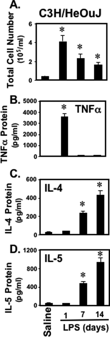
Gram-negative bacterial lipopolysaccharide (LPS) activates macrophages by interacting with Toll-like receptor 4 (TLR4) and triggers the production of various pro-inflammatory Th1 type (type 1) cytokines such as IFNgamma, TNFalpha, and IL8. Though some recent studies cited macrophages as potential sources for Th2 type (type 2) cytokines, little however is known about the intracellular events that lead to LPS-induced type 2 cytokines in macrophages. To understand the mechanisms by which LPS induces type 2 cytokine gene expression, macrophages were stimulated with LPS, and the expression of IL-4 and IL-5 genes were examined. LPS, acting through TLR4, activates both type 1 and type 2 cytokine production both in vitro and in vivo by using macrophages from C3H/HeJ or C3H/HeOuJ mice. Although the baseline level of both TNFalpha and IL-4 protein was very low, TNFalpha was released rapidly after stimulation (within 4 h); however, IL-4 was released after 48 h LPS stimulation in secreted form. Silencing of myeloid differentiation protein (MyD88) and TRIF-related adaptor molecule (TRAM), using small interfering RNA abolished IL-4 induction induced by LPS whereas silencing of TRAM has no effect on TNFalpha induction, thereby indicating that LPS-induced TNFalpha is MyD88-dependent but IL-4 is required both MyD88 and TRAM. These findings suggest a novel function of LPS and the signaling pathways in the induction of IL-4 gene expression.
Figures


Similar articles
-
Melatonin modulates TLR4-mediated inflammatory genes through MyD88- and TRIF-dependent signaling pathways in lipopolysaccharide-stimulated RAW264.7 cells.J Pineal Res. 2012 Nov;53(4):325-34. doi: 10.1111/j.1600-079X.2012.01002.x. Epub 2012 Apr 27. J Pineal Res. 2012. PMID: 22537289
-
The small GTPase Arf6 is essential for the Tram/Trif pathway in TLR4 signaling.J Biol Chem. 2014 Jan 17;289(3):1364-76. doi: 10.1074/jbc.M113.499194. Epub 2013 Dec 2. J Biol Chem. 2014. PMID: 24297182 Free PMC article.
-
TRIF signaling is essential for TLR4-driven IgE class switching.J Immunol. 2014 Mar 15;192(6):2651-8. doi: 10.4049/jimmunol.1300909. Epub 2014 Feb 14. J Immunol. 2014. PMID: 24532577 Free PMC article.
-
IL-21 enhances SOCS gene expression and inhibits LPS-induced cytokine production in human monocyte-derived dendritic cells.J Leukoc Biol. 2006 Jun;79(6):1279-85. doi: 10.1189/jlb.0905503. Epub 2006 Mar 21. J Leukoc Biol. 2006. PMID: 16551679
-
Roles for LPS-dependent interaction and relocation of TLR4 and TRAM in TRIF-signaling.Biochem Biophys Res Commun. 2008 Mar 28;368(1):94-9. doi: 10.1016/j.bbrc.2008.01.061. Epub 2008 Jan 28. Biochem Biophys Res Commun. 2008. PMID: 18222170
Cited by
-
The chemistry of gut microbiome-derived lipopolysaccharides impacts on the occurrence of food allergy in the pediatric age.Front Mol Biosci. 2023 Oct 13;10:1266293. doi: 10.3389/fmolb.2023.1266293. eCollection 2023. Front Mol Biosci. 2023. PMID: 37900913 Free PMC article.
-
BPIFA1 regulates lung neutrophil recruitment and interferon signaling during acute inflammation.Am J Physiol Lung Cell Mol Physiol. 2019 Feb 1;316(2):L321-L333. doi: 10.1152/ajplung.00056.2018. Epub 2018 Nov 21. Am J Physiol Lung Cell Mol Physiol. 2019. PMID: 30461288 Free PMC article.
-
The role of TLR2, TRL3, TRL4, and TRL9 signaling in the pathogenesis of autoimmune disease in a retinal autoimmunity model.Invest Ophthalmol Vis Sci. 2010 Jun;51(6):3092-9. doi: 10.1167/iovs.09-4754. Epub 2010 Jan 27. Invest Ophthalmol Vis Sci. 2010. PMID: 20107166 Free PMC article.
-
LncRNA VINAS regulates atherosclerosis by modulating NF-κB and MAPK signaling.JCI Insight. 2020 Nov 5;5(21):e140627. doi: 10.1172/jci.insight.140627. JCI Insight. 2020. PMID: 33021969 Free PMC article.
-
Lipopolysaccharide endotoxin injections elevated salivary TNFα and corneal temperatures and induced dynamic changes in circulating leukocytes, inflammatory cytokines, and metabolic indicators in wether lambs.J Anim Sci. 2021 Jun 1;99(6):skab120. doi: 10.1093/jas/skab120. J Anim Sci. 2021. PMID: 33871612 Free PMC article.
References
Publication types
MeSH terms
Substances
Grants and funding
LinkOut - more resources
Full Text Sources
Other Literature Sources
