

Diffusion-weighted and Gd-EOB-DTPA-contrast-enhanced magnetic resonance imaging for characterization of tumor necrosis in an animal model
- PMID: 19638862
- PMCID: PMC2771366
- DOI: 10.1097/RCT.0b013e3181953df3
Diffusion-weighted and Gd-EOB-DTPA-contrast-enhanced magnetic resonance imaging for characterization of tumor necrosis in an animal model
Abstract
Purpose: To evaluate the role of diffusion-weighted magnetic resonance imaging (MRI) in determining tumor necrosis and contrast-enhanced MRI using gadoxetic acid disodium (Gd-EOB-DTPA) in determining maximum tumor size measurement and tumor delineation compared with criterion-standard histologic measurements in the rabbit VX2 liver tumor model.
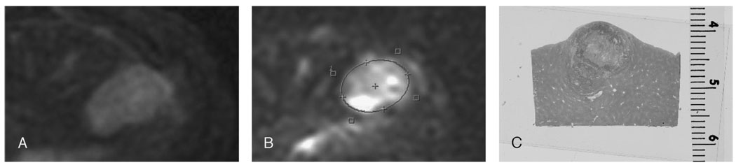
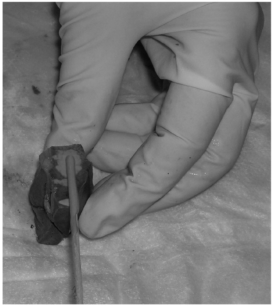
Materials and methods: VX2 tumors were implanted in the livers of 13 rabbits. Magnetic resonance imaging was performed using a 1.5-T MRI scanner and an extremity coil. The imaging protocol included T2-weighted fast spin-echo images, 3-dimensional T1-weighted spoiled gradient-echo with and without fat suppression after administration of Gd-EOB-DTPA, and diffusion-weighted echo planar images. Rabbits were killed, and the tumor was harvested and sliced at 4-mm intervals in the axial plane. The MRI parameters evaluated were tumor size, tumor delineation, and tumor apparent diffusion coefficient (ADC) values. Histologic sections were evaluated to quantify tumor necrosis.
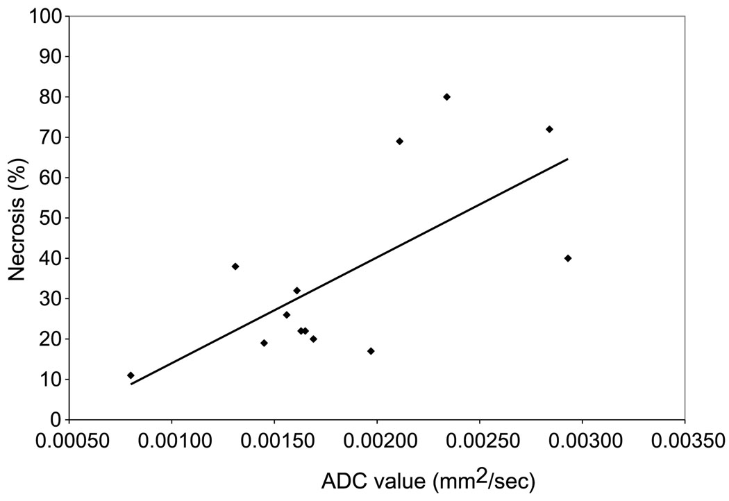
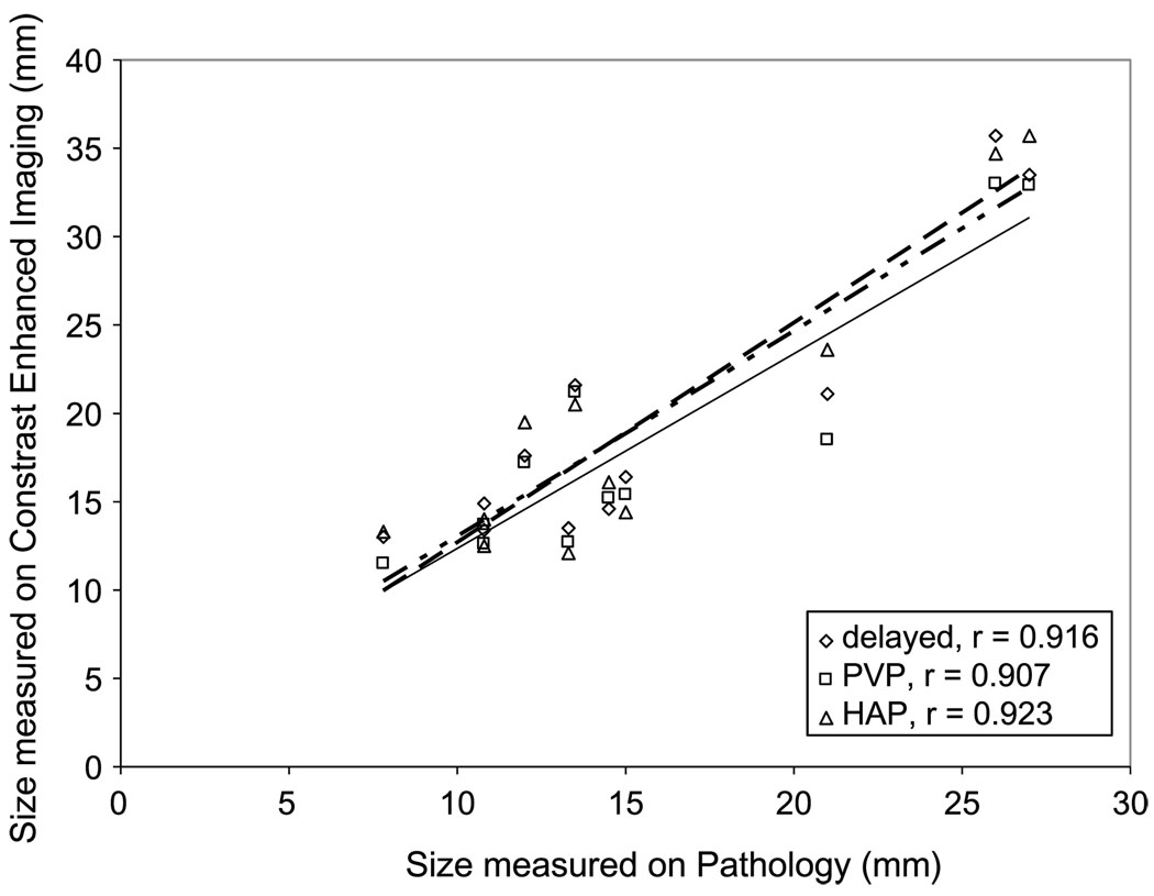
Results: On contrast-enhanced MRI (obtained from 11 rabbits), the mean tumor sizes were 20, 19, and 20 mm in the arterial, portal venous, and delayed phases, respectively. Tumor delineation was most distinguishable in the delayed phase. On diffusion-weighted MRI (acquired in 13 rabbits), the mean tumor ADC value was 1.84 x 10 mm/s. The mean tumor size at pathology was 16 mm. The mean percent necrosis at the tumor's pathologic condition was 36%. The correlation between ADC value and percent necrosis showed an R value of 0.68.
Conclusions: Contrast-enhanced MRI using Gd-EOB-DTPA may provide additional information about tumor outline in the liver. Moreover, we showed a remarkable correlation between ADC values and tumor necrosis. Thus, diffusion-weighted imaging may be useful to assess tumor necrosis; nevertheless, the search for new modalities remains important.
Figures
Similar articles
-
Value of gadoxetic acid-enhanced MRI and diffusion-weighted imaging in the differentiation of hypervascular hyperplastic nodule from small (<3 cm) hypervascular hepatocellular carcinoma in patients with alcoholic liver cirrhosis: A retrospective case-control study.J Magn Reson Imaging. 2020 Jan;51(1):70-80. doi: 10.1002/jmri.26768. Epub 2019 May 6. J Magn Reson Imaging. 2020. PMID: 31062483
-
Prediction of high-stage liver fibrosis using ADC value on diffusion-weighted imaging and quantitative enhancement ratio at the hepatobiliary phase of Gd-EOB-DTPA-enhanced MRI at 1.5 T.Acta Radiol. 2018 May;59(5):509-516. doi: 10.1177/0284185117725778. Epub 2017 Aug 30. Acta Radiol. 2018. PMID: 28853292
-
Diffusion weighted imaging of liver lesions suspect for metastases: Apparent diffusion coefficient (ADC) values and lesion contrast are independent from Gd-EOB-DTPA administration.Eur J Radiol. 2012 Aug;81(8):e849-53. doi: 10.1016/j.ejrad.2012.03.027. Epub 2012 May 8. Eur J Radiol. 2012. PMID: 22575171
-
[Application of Diffusion-Weighted Imaging and Hepatobiliary-Specific Contrast Agent Gd-EOB-DTPA in the Diagnosis and Differential Diagnosis of Focal Liver Lesions].Sichuan Da Xue Xue Bao Yi Xue Ban. 2022 Sep;53(5):737-743. doi: 10.12182/20220960205. Sichuan Da Xue Xue Bao Yi Xue Ban. 2022. PMID: 36224672 Free PMC article. Review. Chinese.
-
Methodology of diffusion-weighted, diffusion tensor and magnetisation transfer imaging.Br J Radiol. 2011 Dec;84 Spec No 2(Spec Iss 2):S121-6. doi: 10.1259/bjr/12789972. Br J Radiol. 2011. PMID: 22433823 Free PMC article. Review.
Cited by
-
Rabbit VX2 Liver Tumor Model: A Review of Clinical, Biology, Histology, and Tumor Microenvironment Characteristics.Front Oncol. 2022 May 10;12:871829. doi: 10.3389/fonc.2022.871829. eCollection 2022. Front Oncol. 2022. PMID: 35619923 Free PMC article. Review.
-
Comparison of gadolinium-EOB-DTPA-enhanced and diffusion-weighted liver MRI for detection of small hepatic metastases.Eur Radiol. 2010 Nov;20(11):2690-8. doi: 10.1007/s00330-010-1842-3. Epub 2010 Jun 20. Eur Radiol. 2010. PMID: 20563726
-
Diffusion-weighted MRI in solitary pulmonary lesions: associations between apparent diffusion coefficient and multiple histopathological parameters.Sci Rep. 2018 Jul 26;8(1):11248. doi: 10.1038/s41598-018-29534-z. Sci Rep. 2018. PMID: 30050167 Free PMC article.
-
Diffusion-weighted imaging of a prostate cancer xenograft model seen on a 7 Tesla animal MR scanner: comparison of ADC values and pathologic findings.Korean J Radiol. 2012 Jan-Feb;13(1):82-9. doi: 10.3348/kjr.2012.13.1.82. Epub 2011 Dec 23. Korean J Radiol. 2012. PMID: 22247640 Free PMC article.
-
Assessment of the hepatic tumor extracellular matrix using elastin-specific molecular magnetic resonance imaging in an experimental rabbit cancer model.Sci Rep. 2020 Nov 27;10(1):20785. doi: 10.1038/s41598-020-77624-8. Sci Rep. 2020. PMID: 33247185 Free PMC article.
References
-
- Vossen JA, Buijs M, Kamel IR. Assessment of tumor response on MR imaging after locoregional therapy. Tech Vasc Interv Radiol. 2006;9:125–132. - PubMed
-
- Le Bihan D, Breton E, Lallemand D, et al. Separation of diffusion and perfusion in intravoxel incoherent motion MR imaging. Radiology. 1988;168:497–505. - PubMed
-
- Benveniste H, Hedlund LW, Johnson GA. Mechanism of detection of acute cerebral ischemia in rats by diffusion-weighted magnetic resonance microscopy. Stroke. 1992;23:746–754. - PubMed
-
- Schaefer PW, Grant PE, Gonzalez RG. Diffusion-weighted MR imaging of the brain. Radiology. 2000;217:331–345. - PubMed
-
- Buijs M, Kamel IR, Vossen JA, et al. Assessment of metastatic breast cancer response to chemoembolization with contrast agent enhanced and diffusion-weighted MR imaging. J Vasc Interv Radiol. 2007;18:957–963. - PubMed
Publication types
MeSH terms
Substances
Grants and funding
LinkOut - more resources
Full Text Sources
Medical
