. 2009 Dec;17(12):2031-40.
doi: 10.1038/mt.2009.174. Epub 2009 Jul 28.

Generating differentially targeted amyloid-beta specific intrabodies as a passive vaccination strategy for Alzheimer's disease

Affiliations

Generating differentially targeted amyloid-beta specific intrabodies as a passive vaccination strategy for Alzheimer's disease

Kelly L Sudol et al. Mol Ther. 2009 Dec.

Abstract

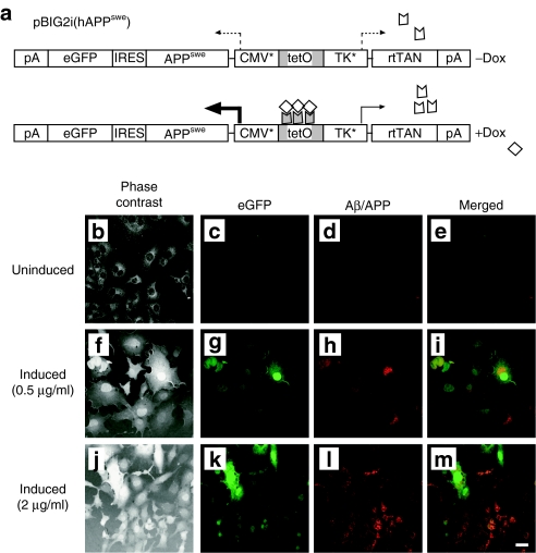
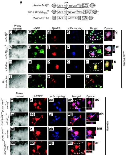
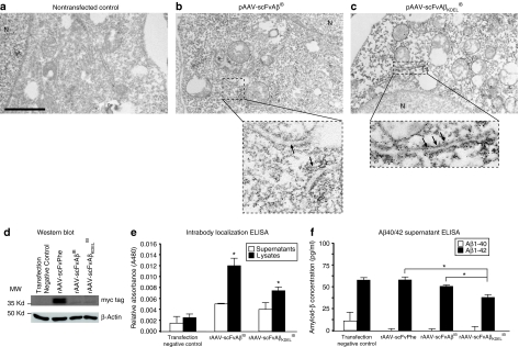
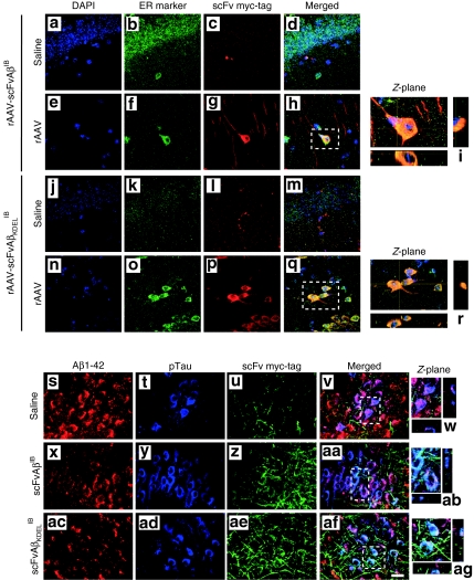
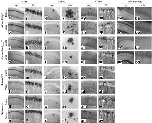
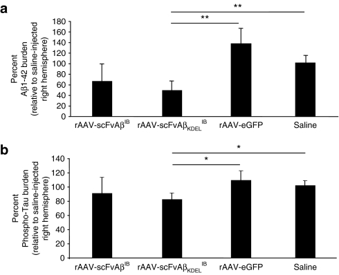
Amyloid-beta (A beta) has been identified as a key component in Alzheimer's disease (AD). Significant in vitro and human pathological data suggest that intraneuronal accumulation of A beta peptides plays an early role in the neurodegenerative cascade. We hypothesized that targeting an antibody-based therapeutic to specifically abrogate intracellular A beta accumulation could prevent or slow disease onset. A beta 42-specific intracellular antibodies (intrabodies) with and without an intracellular trafficking signal were engineered from a previously characterized single-chain variable fragment (scFv) antibody. The intrabodies, one with an endoplasmic reticulum (ER) targeting signal and one devoid of a targeting sequence, were assessed in cells harboring a doxycycline (Dox)-regulated mutant human amyloid precursor protein Swedish mutant (hAPP(swe)) transcription unit for their abilities to prevent A beta peptide egress. Adeno-associated virus (AAV) vectors expressing the engineered intrabodies were administered to young adult 3xTg-AD mice, a model that develops amyloid and Tau pathologies, prior to the initial appearance of intraneuronal A beta. Chronic expression of the ER-targeted intrabody (IB) led to partial clearance of A beta 42 deposits and interestingly, in reduced staining for a pathologic phospho-Tau epitope (Thr231). This approach may provide insights into the functional relevance of intraneuronal A beta accumulation in early AD and potentially lead to the development of new therapeutics.


Figures

<b>Figure 1</b>
Figure 1
Generation of a doxycycline-inducible APPswe stable cell line for characterization of Aβ42-specific intrabodies. (a) To generate a clonal cell line that conditionally expresses the human amyloid precursor protein Swedish mutant (hAPPswe), the hAPPswe gene was inserted into an autoregulated, bidirectional expression vector (pBIG2i14). Following addition of the tetracycline homologue doxycycline (Dox), expression of the chimeric reverse tetracycline transactivator is autoactivated via a Tet operator-controlled synthetic thymidine kinase (TK*) promoter, while a synthetic CMV promoter (CMV*) is simultaneously upregulated to drive expression of the hAPPswe transgene and the downstream reporter gene, enhanced green fluorescent protein (eGFP), via an internal ribosomal entry site. This construct, designated pBIG2i(hAPPswe), was transfected into baby hamster kidney (BHK) cells and placed under hygromycin selection (600 µg/ml). Positive BHK-hAPPswe clones were expanded and coimmunocytochemistry was performed for eGFP and hAPP/amyloid-β (Aβ) on cells incubated in the absence (be) or presence of 0.5 µg/ml Dox (fi) or 2 µg/ml Dox (jm). Cells were visualized using phase contrast (b,f,j) and fluorescence microscopy. Green fluorescence depicts expression from the eGFP reporter gene (c,g,k) and red fluorescence signifies hAPP/Aβ expression (d,h,l). Co-registered staining is indicated in the merged images as yellow (e,i,m). Bar in m = 10 µm. Aβ, amyloid-β.
<b>Figure 2</b>
Figure 2
Colocalization of anti-Aβ42 intrabodies with hAPPswe/Aβ in vitro. (a) Two recombinant adeno-associated virus (rAAV) vectors were constructed: one expressing an Aβ42-specific intrabody sequence with a c-myc epitope tag at the C-terminus to facilitate immunocytochemical detection (rAAV-scFvAβIB), and a second expressing the same c-myc tagged intrabody but with an endoplasmic reticulum targeting signal (KDEL) inserted in-frame between the intrabody coding sequence and c-myc epitope (rAAV-scFvAβKDELIB). The individual transgenes were placed under the transcriptional control of the human cytomegalovirus (CMV) promoter. A polyadenylation signal from SV40 was included at the 3′ end of the transcription unit, which in total was flanked by AAV inverted terminal repeat sequences. A rAAV vector expressing a phenobarbital-specific scFv that had been described previously was used as a negative control for a subset of in vitro studies.45 The rAAV-scFvAβIB (bg), rAAV-scFvAβKDELIB (hm), and rAAV-scFvPhe (ns) plasmids were transiently transfected into the baby hamster kidney (BHK)-human amyloid precursor protein Swedish mutant (hAPPswe) cells incubated in the presence of 2 µg/ml doxycycline (Dox), whereas nontransfected, Dox-treated BHK-hAPPswe cells were used as negative controls (tx). Forty-eight hours post-transfection, coimmunocytochemistry was performed for eGFP (green; c,i,o,u), hAPP/Aβ (red; d,j,p,v), and the c-myc epitope tag (blue; e,k,q,w). Images were obtained by confocal fluorescence microscopy at ×40 original magnification. Co-registered green/red/blue staining is indicated in the merged images as white (f,g,l,m,r,s,x). Co-registered green/red staining is indicated in the merged images as yellow. Panels g,m, and s represent Z-plane images of regions in f,l, and r demarcated with a white dotted box. The rAAV-scFvAβIB (yac), rAAV-scFvAβKDELIB (adah), and rAAV-scFvPhe (aiam) plasmids were also transiently co-transfected with a plasmid expressing hAPPswe into Neuro2A cells with, while Neuro2A cells transfected with only pCMV-hAPPswe were used as controls (anar). Forty-eight hours post-transfection, coimmunocytochemistry was performed for hAPP/Aβ (red; z,ae,aj,ao) and the c-myc epitope tag (blue; aa,af,ak,ap). Images were obtained by confocal fluorescence microscopy at ×40 original magnification. Co-registered red/blue staining is indicated in the merged images as pink (ab,ac,ag,ah,al,am). Panels ac,ah,am, and ar represent Z-plane images of regions in f,l, and r. Bars in x and aq = 10 µm. scFv, single-chain variable fragment; Aβ, amyloid-β; IB, intrabody.
<b>Figure 3</b>
Figure 3
The engineered intrabody constructs maintain Aß42-binding activity, block egress of Aß42, and KDEL-targeted anti-Aß42 intrabody selectively localizes to endoplasmic reticulum ultrastructures within transiently transfected cells. To determine the subcellular localization of the individual intrabodies by immuno-electron microscopy, nontransfected baby hamster kidney (BHK) cells were used as negative controls (a) and the rAAV-scFvAβIB (b) and rAAV-scFvAβKDELib plasmids (c) harboring the anti-Aβ42 intrabody expression cassettes were transiently transfected into BHK cells. Forty-eight hours post-transfection, cell monolayers were fixed and processed for immuno-electron microscopy using a c-myc epitope-specific antibody for detection of the engineered intrabodies. “N” designates the nucleus of the cell. The areas on the ×10,000 photomicrograph demarcated by the dotted boxes were visualized at ×45,000 and included in the respective insets. Arrows point to endoplasmic reticulum ultrastructures. Bar in a = 2,000 nm. (d) The rAAV-scFvAβib and rAAV-scFvAβKDELIB plasmids, as well as the rAAV-scFvPhe control plasmid, were transiently transfected into BHK cells, and 48 hours later, cell lysates were generated and western blot analysis was performed using an anti-myc tag antibody to detect the engineered scFv proteins and an anti-β-actin antibody to assess protein loading. (e) Separately, the rAAV-scFvAβib and rAAV-scFvAβKDELib plasmids were transiently transfected into BHK cells (N = 4). After 48 hours, supernatants (white bars) and cell lysates (black bars) were analyzed by enzyme-linked immunosorbent assay (ELISA) to confirm the intrabodies maintained their ability to bind Aβ42 peptide coated onto microtiter plates. Nontransfected cell supernatants and lysates were used as negative controls. Error bars indicate standard deviation. “*”Indicates P < 0.05 as determined by analysis of variance (ANOVA). (f) The rAAV-scFvAβib and rAAV-scFvAβKDELib plasmids, as well as the rAAV-scFvPhe control plasmid, were transiently transfected into Dox-treated BHK-hAPPswe cells, and 48 hours later, culture supernatants were isolated and ELISA analyses were performed to measure human Aβ40 (white bars) and Aβ42 (black bars) release from the BHK-hAPPswe cells (N = 4). Nontransfected cell supernatants were used as negative controls. Error bars indicate standard deviation. “*”indicates P < 0.05 as determined by ANOVA. scFv, single-chain variable fragment; Aβ, amyloid-β; IB, intrabody; KDEL, lysine-asparticacid-glutanicacid-leucine.
<b>Figure 4</b>
Figure 4
AAV vector–mediated expression of the Aβ-specific intrabodies in 3xTg-AD mice reveals the intrabodies alter general patterns of cell-associated Aβ42. Two month-old 3xTg-AD mice were unilaterally injected in the CA1 region of the hippocampus with saline (ad and jm), recombinant adeno-associated virus (rAAV)-scFvAβIB (ei) and rAAV-scFvAβKDELIB (nr). Nine months postinjection animals were killed and brain sections were processed for coimmunocytochemistry with the following: 4',6-diamidino-2-phenylindole (blue; a,e,j,n), endoplasmic reticulum-specific antibody (green; b,f,k,o) and c-myc epitope tag-specific antibody (red; c,g,l,p). Co-registered staining is indicated in the merged images as yellow (d,h,m,q). Panels i and r represent digitally magnified images, which illustrate a cross section through the Z-plane obtained with confocal microscopy. All images were obtained at ×100 original magnification. Additional brain sections from these rAAV vector-injected 3xTg-AD mice were processed for fluorescence coimmunocytochemistry with the following: Aβ42-specific antibody (red; s,x,ac), phospho-Tau (AT180)-specific antibody (blue; t,y,ad) and c-myc epitope tag-specific antibody (green; u,z,ae). Co-registered staining for all markers is indicated in the merged images as white or for Aβ42 and phospho-Tau as purple (v,w,aa,ab,af,ag). Panels w,ab, and ag represent digitally magnified images, which illustrate a cross section through the Z-plane obtained with confocal microscopy. All images were obtained at ×100 original magnification. scFv, single-chain variable fragment; Aβ, amyloid-β; IB, intrabody.
<b>Figure 5</b>
Figure 5
Qualitative effects of chronic anti-Aβ42 intrabody expression on AD-related pathological hallmarks. Two month-old 3xTg-Alzheimer's disease mice were unilaterally injected in the CA1 region of the hippocampus with saline, recombinant adeno-associated virus (rAAV)-enhanced green fluorescent protein, rAAV-scFvAβIB or rAAV-scFvAβKDELIB. Nine months postinjection animals were killed and 30-µm brain sections were processed for immunohistochemical analyses of hAPP transgene expression (Y188 antibody; ap), extracellular Aβ 1–42 deposition (anti-Aβ42 antibody; qaf), hyperphosphorylated Tau (AT180 antibody; agav), and c-myc epitope tag-specific antibody (awbd). Of note, the images obtained for the c-myc epitope tag immunohistochemical analysis were obtained originally in color by fluorescence microscopy, converted to black-and-white, and subsequently inverted to provide comparable views to the other sections captured using conventional light microscopy following 3,3'-diaminobenzidine immunohistochemistry. Representative images of the infused hippocampus are displayed at ×10 (ah,qx,agan,awaz) and ×40 original magnification (ip,yaf,aoav,babd). scFv, single-chain variable fragment; Aβ, amyloid-β; IB, intrabody.
<b>Figure 6</b>
Figure 6
Recombinant adeno-associated virus (rAAV)-scFvAβKDELIB delivery results in a quantitative reduction in immunoreactive Aβ42 plaque burden and phospho-Tau pathology. Coronal brain sections from 11 month-old 3xTg-Alzheimer's disease (AD) animals that were injected with rAAV-scFvAβIB, rAAV-scFvAβKDELIB, rAAV-enhanced green fluorescent protein, or saline were processed for immunohistochemistry with 3,3'-diaminobenzidine development, and two AD pathologic markers were quantified using the MCID Elite program. The percent of Aβ42 plaque burden relative to the saline-injected right hemisphere (a) was determined by enumerating all the immunoreactive plaque in consecutive coronal brain sections. The percent of phospho-Tau burden was calculated in a similar manner (b). Error bars indicate standard error of the mean. “*” equals P < 0.05 and “**” equals P < 0.001 as determined by Student's t-test. scFv, single-chain variable fragment; Aβ, amyloid-β; IB, intrabody.

Similar articles

Cited by

References

    1. Masters CL, Multhaup G, Simms G, Pottgiesser J, Martins RN., and , Beyreuther K. Neuronal origin of a cerebral amyloid: neurofibrillary tangles of Alzheimer's disease contain the same protein as the amyloid of plaque cores and blood vessels. EMBO J. 1985;4:2757–2763. - PMC - PubMed
    1. Chui DH, Dobo E, Makifuchi T, Akiyama H, Kawakatsu S, Petit A, et al. Apoptotic neurons in Alzheimer's disease frequently show intracellular Abeta42 labeling. J Alzheimers Dis. 2001;3:231–239. - PubMed
    1. Kienlen-Campard P, Miolet S, Tasiaux B., and , Octave JN. Intracellular amyloid-beta 1-42, but not extracellular soluble amyloid-beta peptides, induces neuronal apoptosis. J Biol Chem. 2002;277:15666–15670. - PubMed
    1. LaFerla FM, Tinkle BT, Bieberich CJ, Haudenschild CC., and , Jay G. The Alzheimer's A beta peptide induces neurodegeneration and apoptotic cell death in transgenic mice. Nat Genet. 1995;9:21–30. - PubMed
    1. Takeda K, Araki W., and , Tabira T. Enhanced generation of intracellular Abeta42 amyloid peptide by mutation of presenilins PS1 and PS2. Eur J Neurosci. 2004;19:258–264. - PubMed

Publication types

MeSH terms