

Clinical Trial
doi: 10.1038/mt.2009.169.
Epub 2009 Jul 28.
Phase I study of H5.020CMV.PDGF-beta to treat venous leg ulcer disease
Affiliations
- PMID: 19638959
- PMCID: PMC2835007
- DOI: 10.1038/mt.2009.169
Item in Clipboard
Clinical Trial
Phase I study of H5.020CMV.PDGF-beta to treat venous leg ulcer disease
Mol Ther.
2009 Oct.
Abstract
Venous leg ulcers are a prevalent nonhealing wound of the lower extremity. Although topically applied growth factors successfully improve wound repair in animal studies, similar studies on humans with venous leg ulcers have not been successful. This study was designed to evaluate the acute safety and biologic feasibility of peri-ulcer injection of a replication-incompetent adenoviral construct expressing platelet-derived growth factor-beta (PDGF-beta). In this phase I study, we demonstrate the initial safety, feasibility, and biologic plausibility of using H5.020CMV.PDGF-beta to treat venous leg ulcer disease.
Figures
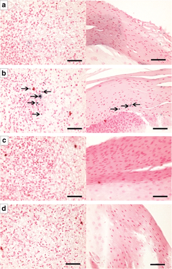
DNA in situ hybridization for the cytomegalovirus (CMV) promoter. (a) Day 1 venous leg ulcer biopsy 5-µm sections showing no CMV-expressing cells in the wound bed (left panel) or in the keratinocyte layer (right panel). (b) Day 3 venous leg ulcer 5-µm sections showing CMV-expressing cells (arrows) in both the wound bed and keratinocyte layer. (c) Day 28 venous leg ulcer 5-µm section showing no CMV-expressing cells, suggesting that the infection has been cleared. (d) Day 3 no probe control venous leg ulcer 5-µm sections showing no positive staining. Bar = 50 µm. Samples were counterstained with Nuclear Fast Red (Vector Laboratories).
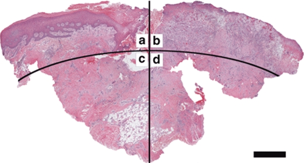
Representative image of a wound biopsy after hematoxylin and eosin staining showing the regions used for cell quantitation and vessel density analysis. (a) Superficial subepithelial region, (b) superficial ulcer region, (c) subepithelial dermal (or deep) region, and (d) ulcer deep region. Bar = 1 mm.
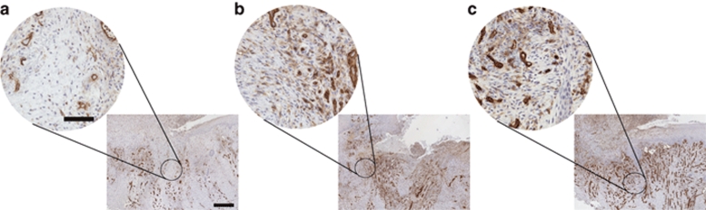
To assess wound neovascularization after administration of H5.020CMV.PDGF-β, we immunostained tissue biopsies for an endothelial specific antigen CD31 and quantified the number of positive staining vessels per HPF at (a) day 1, (b) day 3, and (c) day 28. Bar = low power: 500 µm, high power: 150 µm. CMV, cytomegalovirus; HPF, high power field; PDGF-β, platelet-derived growth factor-β.
To assess wound inflammation from H5.020CMV.PDGF-β, we immunostained tissue biopsies for the common leukocyte antigen (CD45) and quantified the number of cells staining positive at (a) day 1, (b) day 3, and (c) day 28. No significant increase in CD45+ cells were noted in tissue biopsies taken at day 1, day 3 and day 28 (P > 0.30). Bar = low power: 500 µm, high power: 50 µm. CMV, cytomegalovirus; PDGF-β, platelet-derived growth factor-β.
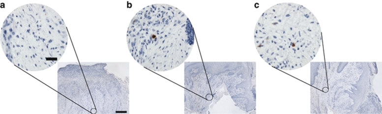
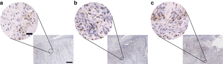
To determine the effect of recruitment on progenitor cells within the wound after administration of H5.020CMV.PDGF-β, we immunostained tissue biopsies for CD133 an antigen found on undifferentiated cell types such as endothelial progenitor cells. High and low power views of representative wound biopsies at (a) day 1, (b) day 3, and (c) day 28. Note more cells staining positive for CD133 at day 3 as compared to day 1 (P = 0.0015). Bar = low power: 500 µm, high power: 50 µm. CMV, cytomegalovirus; PDGF-β, platelet-derived growth factor-β.
References
-
- Ruckley CV. Socioeconomic impact of chronic venous insufficiency and leg ulcers. Angiology. 1997;48:67–69. - PubMed
-
- Bickers DR, Lim HW, Margolis D, Weinstock MA, Goodman C, Faulkner E, et al. The burden of skin diseases: 2004 a joint project of the American Academy of Dermatology Association and the Society for Investigative Dermatology. J Am Acad Dermatol. 2006;55:490–500. - PubMed
-
- Coon WW, Willis PW., and , Keller JB. Venous thromboembolism and other venous disease in the Tecumseh community health study. Circulation. 1973;48:839–846. - PubMed
-
- Hallbook T. Leg ulcer epidemiology. Acta Chir Scand. 1988;544:17–20. - PubMed
-
- Phillips TJ. Chronic cutaneous ulcers: etiology and epidemiology. J Invest Dermatol. 1994;102:38S–41S. - PubMed
Publication types
MeSH terms
Substances
Grants and funding
LinkOut - more resources
Full Text Sources
Medical
