. 2009:2009:619487.
doi: 10.1155/2009/619487. Epub 2009 Jul 21.

Pyrrolidine dithiocarbamate attenuates paraquat-induced lung injury in rats

Affiliations

Pyrrolidine dithiocarbamate attenuates paraquat-induced lung injury in rats

Xiuli Chang et al. J Biomed Biotechnol. 2009.

Abstract

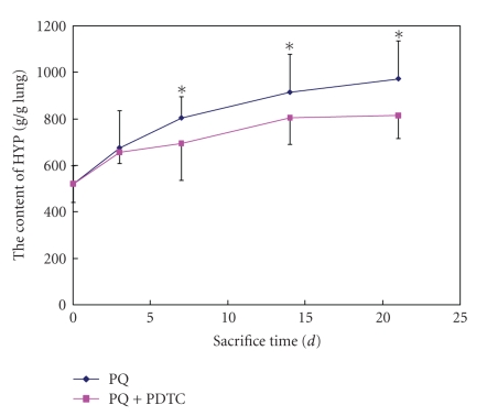
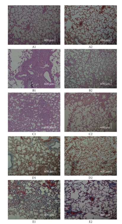
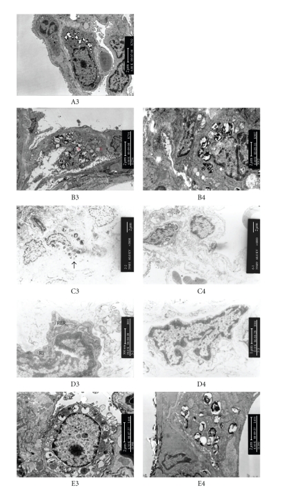
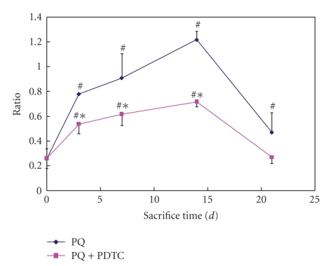
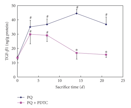
Paraquat (PQ) has been demonstrated that the main target organ for the toxicity is the lung. This study aimed to investigate the potential protective effect of PDTC on the PQ-induced pulmonary damage. Fifty-four rats were divided into control, PQ-treated and PQ+PDTC-treated groups. Rats in the PQ group were administrated 40 mg/kg PQ by gastric gavage, and PDTC group with 40 mg/kg PQ followed by injection of 120 mg/kg PDTC (IP). On the days 3, 7, 14 and 21 after treatments, the activities of GSH-Px, SOD, MDA level and the content of HYP were measured. TGF-beta1 mRNA and protein were assayed by RT-PCR and ELISA. MDA level in plasma and BALF was increased and the activities of GSH-Px and SOD were decreased significantly in the PQ-treated groups (P < .05) compared with control group. While the activities of GSH-Px and SOD in the PQ+PDTC-treated groups was markedly higher than that of PQ-treated groups (P < .05), and in contrast, MDA level was lower. TGF-beta1 mRNA and protein were significantly lower in the PQ+PDTC-treated groups than that of PQ-treated groups (P < .05). The histopathological changes in the PQ+PDTC-treated groups were milder than those of PQ groups. Our results suggested that PDTC treatment significantly attenuated paraquat-induced pulmonary damage.


Figures

Figure 1
Figure 1
The content of HYP in lung tissue from groups of the control, PQ treatment, and PQ+PDTC treatment. *compared with the control, P < .01
Figure 2
Figure 2
The left lung histological slices of SD rats from groups of the control, PQ treatment, and PQ+PDTC treatment. In A1 (light micrographs of lung sections stained with hematoxylin and eosin (HE) from control), evidencing a normal pulmonary structure without evidence of alveolar collapse, vascular congestion, or cellular infiltrations. In B1(PQ group on the 3rd day HE), the capillary vessel of alveolar wall dilation, congestion in lungs, hemorrhagic lung, and alveolar epithelium detached, edema, and infiltration of inflammatory cell around bronchia can be visible. In B2 (PQ+PDTC group on the 3rd day HE), a slight decrease in the alveolar space can be observed as well as the existence of several infiltrative vacuolated cells in the interstitial and in the alveolar space. In C1 (PQ group on the 7th day HE), the lungs showed an accumulation of mixed inflammatory cells in the alveolar region and confluent areas of marked interstitial thickening. In C2 (PQ+PDTC group on the 7th day HE), there was a slight change. In A2 (MASSON from control group), relatively little collagen accumulation can be observed in control group. In D1 (PQ group on the 14th day MASSON) and E1 (PQ group on the 21st day MASSON), Masson's Trichrome staining showed collagen accumulation, broadened alveolar wall and patchy collagen deposition within the expanded interstitium in some areas. In D2 (PQ+PDTC group on the 14th day MASSON) and E2 (PQ+PDTC group on the 21st day MASSON), a slight collagen accumulation can be visible.
Figure 3
Figure 3
The lung ultrastructure of SD rats from groups of the control, PQ treatment, and PQ+PDTC treatment. Electron micrographs from control group (A3) showed a normal ultrastructure with the presence of pneumocytes type I and II. B3 (PQ group on the 3rd day) and C3 (PQ group on the 7th day) can be observed a type II pneumocyte with mitochondrial swelling (b), dilation of lamellar body (a), pneumocyte necrosis and alveolar collapse (↑). B4 (PQ+PDTC group on the 3rd day) and C4 (PQ+PDTC group on the 7th day) can be observed normal mitochondria and no alveolar collapse. D3 (PQ group on the 14th day) can be noticed the more numerous rough endroplasmic reticulum (RER) and ribosome (Ri) in fibroblast compared with D4 (PQ+PDTC group on the 14th day). showed E3 (PQ group on the 21st day) can be observed patchy collagen deposition within the expanded interstitium in some areas. E4 (PQ+PDTC group on the 21st day) can be observed a small quantity of collagen deposition.
Figure 4
Figure 4
Expression TGF-β1 mRNA in lung tissue from groups of the control, PQ treatment, and PQ+PDTC treatment. Relative TGF-β1 mRNA abundance was normalized with the β-actin, and expressed as relative units. Values are given as mean ± SE (n = 3). # Compared with the control, P < .05; *compared with the PQ treatment, P < .05.
Figure 5
Figure 5
TGF-β1 level in plasm at sacrifice time from groups of the control, PQ treatment, and PQ+PDTC treatment. TGF-β1 was expressed as ng/g of protein. Values are given as mean ± SE (n = 3). # Compared with the control, P < .05; *compared with the PQ treatment, P < .05.

References

    1. Dinis-Oliveira RJ, De Jesús Valle MJ, Bastos ML, Carvalho F, Sánchez Navarro A. Kinetics of paraquat in the isolated rat lung: influence of sodium depletion. Xenobiotica. 2006;36(8):724–737. - PubMed
    1. Dinis-Oliveira RJ, Remião F, Carmo H, et al. Paraquat exposure as an etiological factor of Parkinson's disease. NeuroToxicology. 2006;27(6):1110–1122. - PubMed
    1. Lewis CP, Nemery B. Pathophysiology and Biochemical Mechanisms of the Pulmonary Toxicity of Paraquat. Vol. 10. New York, NY, USA: Marcel Dekker; 1995.
    1. Gil HW, Oh MH, Woo KM, Lee EY, Oh MH, Hong SY. Relationship between pulmonary surfactant protein and lipid peroxidation in lung injury due to paraquat intoxication in rats. The Korean Journal of Internal Medicine. 2007;22(2):67–72. - PMC - PubMed
    1. Yamashita M, Yamashita M, Ando Y. A long-term follow-up of lung function in survivors of paraquat poisoning. Human and Experimental Toxicology. 2000;19(2):99–103. - PubMed

Publication types

LinkOut - more resources