Exploring the structure of the N-terminal domain of CP29 with ultrafast fluorescence spectroscopy
- PMID: 19639311
- PMCID: PMC2841283
- DOI: 10.1007/s00249-009-0519-9
Exploring the structure of the N-terminal domain of CP29 with ultrafast fluorescence spectroscopy
Abstract
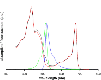
A high-throughput Förster resonance energy transfer (FRET) study was performed on the approximately 100 amino acids long N-terminal domain of the photosynthetic complex CP29 of higher plants. For this purpose, CP29 was singly mutated along its N-terminal domain, replacing one-by-one native amino acids by a cysteine, which was labeled with a BODIPY fluorescent probe, and reconstituted with the natural pigments of CP9, chlorophylls and xanthophylls. Picosecond fluorescence experiments revealed rapid energy transfer (approximately 20-70 ps) from BODIPY at amino-acid positions 4, 22, 33, 40, 56, 65, 74, 90, and 97 to Chl a molecules in the hydrophobic part of the protein. From the energy transfer times, distances were estimated between label and chlorophyll molecules, using the Förster equation. When the label was attached to amino acids 4, 56, and 97, it was found to be located very close to the protein core (approximately 15 A), whereas labels at positions 15, 22, 33, 40, 65, 74, and 90 were found at somewhat larger distances. It is concluded that the entire N-terminal domain is in close contact with the hydrophobic core and that there is no loop sticking out into the stroma. Most of the results support a recently proposed topological model for the N-terminus of CP29, which was based on electron-spin-resonance measurements on spin-labeled CP29 with and without its natural pigment content. The present results lead to a slight refinement of that model.
Figures


Similar articles
-
Site-directed spin-labeling study of the light-harvesting complex CP29.Biophys J. 2009 May 6;96(9):3620-8. doi: 10.1016/j.bpj.2009.01.038. Biophys J. 2009. PMID: 19413967 Free PMC article.
-
Spectroscopic study of the light-harvesting CP29 antenna complex of photosystem II--part I.J Phys Chem B. 2013 Jun 6;117(22):6585-92. doi: 10.1021/jp4004328. Epub 2013 May 21. J Phys Chem B. 2013. PMID: 23631672
-
Modeling of optical spectra of the light-harvesting CP29 antenna complex of photosystem II--part II.J Phys Chem B. 2013 Jun 6;117(22):6593-602. doi: 10.1021/jp4004278. Epub 2013 May 21. J Phys Chem B. 2013. PMID: 23662835
-
Structural features of the diatom photosystem II-light-harvesting antenna complex.FEBS J. 2020 Jun;287(11):2191-2200. doi: 10.1111/febs.15183. Epub 2020 Jan 7. FEBS J. 2020. PMID: 31854056 Review.
-
The significance of CP29 reversible phosphorylation in thylakoids of higher plants under environmental stresses.J Exp Bot. 2013 Mar;64(5):1167-78. doi: 10.1093/jxb/ert002. Epub 2013 Jan 23. J Exp Bot. 2013. PMID: 23349136 Review.
Cited by
-
The origin of pigment-binding differences in CP29 and LHCII: the role of protein structure and dynamics.Photochem Photobiol Sci. 2023 Jun;22(6):1279-1297. doi: 10.1007/s43630-023-00368-7. Epub 2023 Feb 6. Photochem Photobiol Sci. 2023. PMID: 36740636
-
Exploring the structure of the 100 amino-acid residue long N-terminus of the plant antenna protein CP29.Biophys J. 2014 Mar 18;106(6):1349-58. doi: 10.1016/j.bpj.2013.11.4506. Biophys J. 2014. PMID: 24655510 Free PMC article.
References
Publication types
MeSH terms
Substances
LinkOut - more resources
Full Text Sources

