. 2009 Dec;118(6):723-36.
doi: 10.1007/s00412-009-0230-8. Epub 2009 Jul 29.

Meiotic behavior of aneuploid chromatin in mouse models of Down syndrome

Affiliations

Meiotic behavior of aneuploid chromatin in mouse models of Down syndrome

Laura G Reinholdt et al. Chromosoma. 2009 Dec.

Abstract

Aneuploidy, which leads to unpaired chromosomal axes during meiosis, is frequently accompanied by infertility. We previously showed, using three mouse models of Down syndrome, that it is an extra chromosome, but not extra gene dose, that is associated with male infertility and virtual absence of post-meiotic gem cells. Here, we test the hypothesis that aneuploid segments are differentially modified and expressed during meiosis, depending on whether they are present as an extra chromosome or not. In all three models examined, the trisomic region lacks a pairing partner, but in one case, spermatocytes have an extra (and unpaired) chromosome, while the two other models involve translocation of the trisomic region rather than an extra chromosome. An extra unpaired chromosome was always modified by phosphorylation of histone H2AX and lacked RNA PolII. But in the case of trisomic regions attached to a paired chromosome, assembly of these protein modifications was affected by the position of a trisomic region relative to a centromere and the physical extent of the unpaired chromatin. Analysis of gene expression in testes revealed that extra copy number alone was not sufficient for meiotic upregulation of genes in the trisomic interval. Additionally and unexpectedly, presence of meiotic gene silencing chromatin modifications was not sufficient for downregulation of genes in unpaired trisomic chromatin. Thus, the meiotic chromatin modifications that are cytologically visible are unlikely to be directly involved in sterility versus fertility of DS models. Finally, the presence of an extra unpaired chromosome, but not the presence of extra (trisomic) genes, caused global deregulation of transcription in spermatocytes. These results reveal mechanisms by which an extra chromosome, but not trisomic gene dose, impact on meiotic progress and infertility.


Figures

Figure 1
Figure 1
Diagrammatic representation of trisomy in three Down syndrome mouse models: Ts(1716)65Dn (Ts65Dn), Rb(12.T171665Dn)2Cje (Rb2Cje) and Ts(16C-tel)1Cje (Ts1Cje). Each of these models is trisomic for the portion of mouse Chr 16 that shares conserved synteny with human Chr 21. Chr 12 and the trisomic segments of Chr 16 are shown in black and red, respectively. Blue represents the Chr 17 centromere. Ts65Dn mice have a small (13.4 Mb) supernumary chromosome that is the product of a translocation between the Chr 17 centromere and the distal end of Chr 16, which shares conserved synteny with human Chr 21. Rb2Cje mice carry the product of a Robertsonian fusion between the small Ts65 chromosome and the centromeric end of Chr 12. Ts1Cje mice carry the product of a serendipitous fusion between a smaller region (compared to the Ts65 chromosome) of distal Chr 16 (8 Mb), with the distal end of Chr 12.
Figure 2
Figure 2
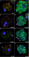
Localization of γH2AX (blue) and RNA POLII (green) in pachytene stage spermatocytes from trisomy mice (Ts65Dn and Ts1Cje) and from diploid littermates (WT). DNA FISH with a Chr 16 BAC probe (red) was used to visualize Chr 16, the Ts65 chromosome and the trisomic Chr 16 segment (16C-tel) in Ts1Cje. In Ts65Dn spermatocytes, the Ts65 chromosome is always associated with the Chr 16 bivalent (E, arrows) and/ or with the XY body (C, D, F). In diploid pachytene spermatocytes, γH2AX (blue) specifically localizes to the XY body (A). In Ts65Dn pachytene spermatocytes, γH2AX (blue) localizes to the Ts65 chromosome whether or not the Ts65 chromosome is associated with the sex body (C, E). In diploid pachytene spermatocytes, the XY body and the chromocenters are depleted of RNA POLII (green, B). The Ts65 chromosome is also depleted of RNA POLII (green, D, E). In Ts1Cje spermatocytes, γH2AX (blue) does not associate with the trisomic segment of Chr 16 (G) and the trisomic segment is not depleted of RNA POLII (green, H). 100X objective.
Figure 3
Figure 3
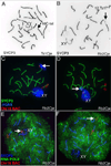
Modification of the unpaired, trisomic segment of Chr 16 in Rb2Cje spermatocytes. Inverted anti-SYCP3 images of a Ts1Cje and Rb2Cje spermatocyte nuclei (A, B respectively). The unpaired segment (16C-tel) in the Ts1Cje spermatocyte nucleus is not visible by standard light microsopy (DNA FISH was used to identify 16C-tel in this spermatocyte) (A). In contrast, the unpaired segment (Ts1716) in an Rb2Cje spermatocyte nucleus is visible (B). The unpaired segment in Rb2Cje spermatocytes is modified by γH2AX regardless of its association with the sex body (C, D, arrows) and is associated with RNA Pol II only when it is not associated with the sex body (E, F, arrows). 100X objective.
Figure 4
Figure 4
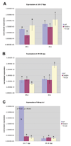
Quantitative RT-PCR profiling of Mlh1 Wrb (A, B) and Rbmy1a1 (C) in enriched spermatocyte preparations from diploid and trisomy testes. The normalized expression value of each gene in Ts65Dn, Ts1Cje and diploid spermatocytes (WT) is plotted directly. The expression was determined for each gene in triplicate samples for each of three biological replicates for each genotype and time point. Values represented here are mean values from triplicate samples from one representative biological replicate. Mean ± SD (n = 3 experimental replicates). Bars within a panel marked with the same letter represent expression values that are not significantly different, while those marked with different letters are different represent expression values that are significantly different (p< 0.05).
Figure 5
Figure 5
Expression of genes within the Ts65Dn trisomy interval (as defined by (Kahlem et al. 2004); Chr 16: ∼84,700,000 bp – 98,100,000 bp; or Mrpl39Znf295) that showed significant (q≤ 0.1) differential expression compared to that of wild type, diploid (WT) enriched spermatocyte preparations from 19–20 dpp testes.
Figure 6
Figure 6
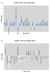
Expression of all genes that showed differential expression (q≤ 0.1) in Ts1Cje enriched spermatocyte preparations compared to that from wild type, diploid (WT) littermates at 16–17 dpp and 19–20 dpp

References

    1. Baarends WM, Wassenaar E, van der Laan R, Hoogerbrugge J, Sleddens-Linkels E, Hoeijmakers JH, de Boer P, Grootegoed JA. Silencing of unpaired chromatin and histone H2A ubiquitination in mammalian meiosis. Mol Cell Biol. 2005;25:1041–1053. - PMC - PubMed
    1. Barlow AL, Benson FE, West SC, Hulten MA. Distribution of the RAD51 recombinase in human and mouse spermatocytes. EMBO J. 1997;16:5207–5215. - PMC - PubMed
    1. Bustin SA. Quantification of mRNA using real-time reverse transcription PCR (RT-PCR): Trends and problems. J Mol Endocrinol. 2002;29:23–39. - PubMed
    1. Cheng Z, Tu C, Rodriguez L, Chen TH, Dvorak MM, Margeta M, Gassmann M, Bettler B, Shoback D, Chang W. Type B gamma-aminobutyric acid receptors modulate the function of the extracellular Ca2+sensing receptor and cell differentiation in murine growth plate chondrocytes. Endocrinology. 2007;148:4984–4992. - PubMed
    1. Cui X, Hwang JT, Qiu J, Blades NJ, Churchill GA. Improved statistical tests for differential gene expression by shrinking variance components estimates. Biostatistics. 2005;6:59–75. - PubMed

Publication types

MeSH terms