

The development of the rat model of retinopathy of prematurity
- PMID: 19639356
- PMCID: PMC3752888
- DOI: 10.1007/s10633-009-9180-y
The development of the rat model of retinopathy of prematurity
Abstract
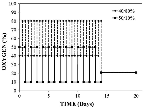
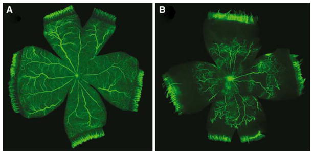
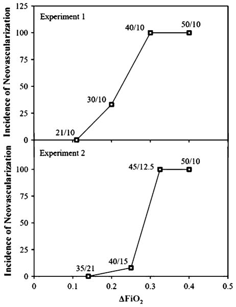
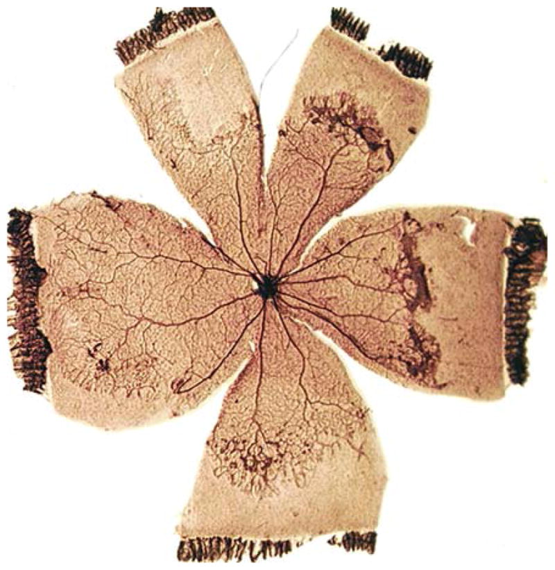
Retinopathy of prematurity (ROP) is a potentially blinding disease affecting premature infants. ROP is characterized by pathological ocular angiogenesis or retinal neovascularization (NV). Models of ROP have yielded much of what is currently known about physiological and pathological blood vessel growth in the retina. The rat provides a particularly attractive and cost effective model of ROP. The rat model of ROP consistently produces a robust pattern of NV, similar to that seen in humans. This model has been used to study gross aspects of angiogenesis. More recently, it has been used to identify and therapeutically target specific genes and molecular mechanisms involved in the angiogenic cascade. As angiogenesis occurs as a complication of many diseases, knowledge gained from these studies has the potential to impact nonocular angiogenic conditions. This article provides historical perspective on the development and use of the rat model of ROP. Key findings generated through the use of this model are also summarized.
Figures
References
-
- Terry TL. Extreme prematurity and fibroblastic overgrowth of persistent vascular sheath behind each crystalline lens: I, preliminary report. Am J Ophthalmol. 1942;25:203–204. - PubMed
-
- Campbell K. Intensive oxygen therapy as a possible cause of retrolental fibroplasia; a clinical approach. Med J Aust. 1951;2:48–50. - PubMed
-
- Patz A, Hoech LE, Cruz ED. Studies on the effect of high oxygen administration in retrolental fibroplasia. I. Nursery observations. Am J Ophthalmol. 1953;35:1245–1253. - PubMed
-
- Kinsey VE. Retrolental fibroplasia; cooperative study of retrolental fibroplasia and the use of oxygen. AMA Arch Ophthalmol. 1956;56:481–543. - PubMed
-
- Lucey JF, Dangman B. A reexamination of the role of oxygen in retrolental fibroplasia. Pediatrics. 1984;73:82–96. - PubMed
Publication types
MeSH terms
Substances
Grants and funding
LinkOut - more resources
Full Text Sources
