

Specialization in the default mode: Task-induced brain deactivations dissociate between visual working memory and attention
- PMID: 19639552
- PMCID: PMC6870780
- DOI: 10.1002/hbm.20850
Specialization in the default mode: Task-induced brain deactivations dissociate between visual working memory and attention
Abstract
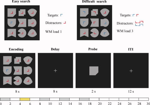
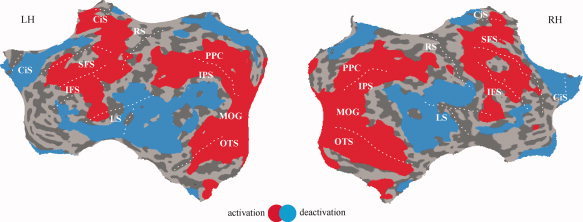
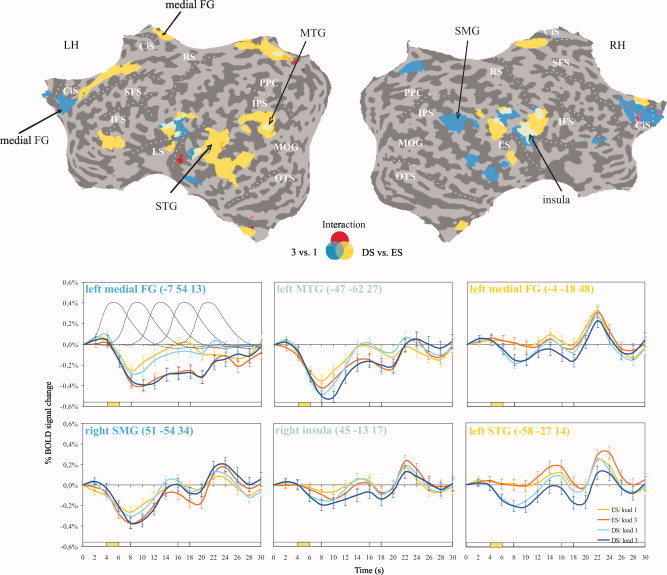
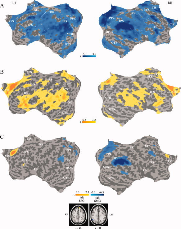
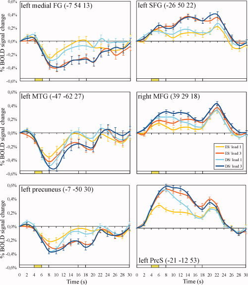
The idea of an organized mode of brain function that is present as default state and suspended during goal-directed behaviors has recently gained much interest in the study of human brain function. The default mode hypothesis is based on the repeated observation that certain brain areas show task-induced deactivations across a wide range of cognitive tasks. In this event-related functional resonance imaging study we tested the default mode hypothesis by comparing common and selective patterns of BOLD deactivation in response to the demands on visual attention and working memory (WM) that were independently modulated within one task. The results revealed task-induced deactivations within regions of the default mode network (DMN) with a segregation of areas that were additively deactivated by an increase in the demands on both attention and WM, and areas that were selectively deactivated by either high attentional demand or WM load. Attention-selective deactivations appeared in the left ventrolateral and medial prefrontal cortex and the left lateral temporal cortex. Conversely, WM-selective deactivations were found predominantly in the right hemisphere including the medial-parietal, the lateral temporo-parietal, and the medial prefrontal cortex. Moreover, during WM encoding deactivated regions showed task-specific functional connectivity. These findings demonstrate that task-induced deactivations within parts of the DMN depend on the specific characteristics of the attention and WM components of the task. The DMN can thus be subdivided into a set of brain regions that deactivate indiscriminately in response to cognitive demand ("the core DMN") and a part whose deactivation depends on the specific task.
2009 Wiley-Liss, Inc.
Figures
Similar articles
-
Common deactivation patterns during working memory and visual attention tasks: an intra-subject fMRI study at 4 Tesla.Hum Brain Mapp. 2006 Aug;27(8):694-705. doi: 10.1002/hbm.20211. Hum Brain Mapp. 2006. PMID: 16404736 Free PMC article.
-
Shared and differential default-mode related patterns of activity in an autobiographical, a self-referential and an attentional task.PLoS One. 2019 Jan 4;14(1):e0209376. doi: 10.1371/journal.pone.0209376. eCollection 2019. PLoS One. 2019. PMID: 30608970 Free PMC article.
-
Coactivation of the Default Mode Network regions and Working Memory Network regions during task preparation.Sci Rep. 2014 Aug 5;4:5954. doi: 10.1038/srep05954. Sci Rep. 2014. PMID: 25092432 Free PMC article.
-
Neuroimaging studies of working memory: a meta-analysis.Cogn Affect Behav Neurosci. 2003 Dec;3(4):255-74. doi: 10.3758/cabn.3.4.255. Cogn Affect Behav Neurosci. 2003. PMID: 15040547 Review.
-
Cortico-cerebellar networks for visual attention and working memory.Curr Opin Psychol. 2019 Oct;29:239-247. doi: 10.1016/j.copsyc.2019.05.003. Epub 2019 May 21. Curr Opin Psychol. 2019. PMID: 31202085 Free PMC article. Review.
Cited by
-
Stress Impact on Resting State Brain Networks.PLoS One. 2013 Jun 19;8(6):e66500. doi: 10.1371/journal.pone.0066500. Print 2013. PLoS One. 2013. PMID: 23840493 Free PMC article.
-
Altered Vision-Related Resting-State Activity in Pituitary Adenoma Patients with Visual Damage.PLoS One. 2016 Aug 11;11(8):e0160119. doi: 10.1371/journal.pone.0160119. eCollection 2016. PLoS One. 2016. PMID: 27512990 Free PMC article.
-
Fluid intelligence allows flexible recruitment of the parieto-frontal network in analogical reasoning.Front Hum Neurosci. 2011 Mar 1;5:22. doi: 10.3389/fnhum.2011.00022. eCollection 2011. Front Hum Neurosci. 2011. PMID: 21415916 Free PMC article.
-
EEG microstates associated with intra- and inter-subject alpha variability.Sci Rep. 2020 Feb 12;10(1):2469. doi: 10.1038/s41598-020-58787-w. Sci Rep. 2020. PMID: 32051420 Free PMC article.
-
Dynamic causal brain circuits during working memory and their functional controllability.Nat Commun. 2021 Jun 29;12(1):3314. doi: 10.1038/s41467-021-23509-x. Nat Commun. 2021. PMID: 34188024 Free PMC article.
References
-
- Amedi A, Malach R, Pascual‐Leone A ( 2005): Negative BOLD differentiates visual imagery and perception. Neuron 48: 859–872. - PubMed
-
- Bar M, Aminoff E, Mason M, Fenske M ( 2007): The units of thought. Hippocampus 17: 420–428. - PubMed
-
- Binder JR, Frost JA, Hammeke TA, Bellgowan PSF, Rao SM, Cox RW ( 1999): Conceptual processing during the conscious resting state. A functional MRI study. J Cogn Neurosci 11: 80–95. - PubMed
-
- Biswal B, Yetkin FZ, Haughton VM, Hyde JS ( 1995): Functional connectivity in the motor cortex of resting human brain using echo‐planar MRI. Magn Reson Med 34: 537–541. - PubMed
Publication types
MeSH terms
Grants and funding
LinkOut - more resources
Full Text Sources
