. 2010 Feb;31(2):222-36.
doi: 10.1002/hbm.20859.

Neural correlates of efficacy of voice therapy in Parkinson's disease identified by performance-correlation analysis

Affiliations

Neural correlates of efficacy of voice therapy in Parkinson's disease identified by performance-correlation analysis

Shalini Narayana et al. Hum Brain Mapp. 2010 Feb.

Abstract

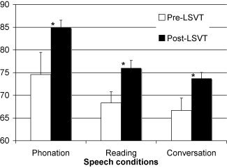
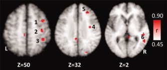
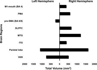
LSVT LOUD (Lee Silverman Voice Treatment) is efficacious in the treatment of speech disorders in idiopathic Parkinson's disease (IPD), particularly hypophonia. Functional imaging in patients with IPD has shown abnormalities in several speech regions and changes in these areas immediately following treatment. This study serves to extend the analysis by correlating changes of regional neural activity with the main behavioral change following treatment, namely, increased vocal intensity. Ten IPD participants with hypophonia were studied before and after LSVT LOUD. Cerebral blood flow during rest and reading conditions were measured by H(2)(15)O-positron emission tomography. Z-score images were generated by contrasting reading with rest conditions for pre- and post-LSVT LOUD sessions. Neuronal activity during reading in the pre- versus post-LSVT LOUD contrast was correlated with corresponding change in vocal intensity to generate correlation images. Behaviorally, vocal intensity for speech tasks increased significantly after LSVT LOUD. The contrast and correlation analyses indicate a treatment-dependent shift to the right hemisphere with modification in the speech motor regions as well as in prefrontal and temporal areas. We interpret the modification of activity in these regions to be a top-down effect of LSVT LOUD. The absence of an effect of LSVT LOUD on the basal ganglion supports this argument. Our findings indicate that the therapeutic effect of LSVT LOUD in IPD hypophonia results from a shift in cortical activity to the right hemisphere. These findings demonstrate that the short-term changes in the speech motor and multimodal integration areas can occur in a top-down manner.


Figures

Figure 1
Figure 1
Behavioral data: changes in measure of loudness as sound pressure level (SPL) in decibels (dB) during sustained phonation, reading, and conversation pre and post‐LSVT LOUD. *Post‐LSVT LOUD loudness was significantly different (P < 0.0005).
Figure 2
Figure 2
Activation pattern during paragraph reading in individuals with IPD hypophonia. Top panel A: Pre LSVT LOUD and bottom panel B: Post‐LSVT LOUD. (1) Bilateral SMA, (2) right PMd, (3) left primary motor cortex (M1‐mouth), (4) right primary motor cortex (M1‐mouth), (5) right parietal cortex (BA 7), (6) right dorsolateral prefrontal cortex (BA 9), and (7) right superior temporal cortex. The figures in the last column are coronal sections (at x = 52) show increased right M1 activation, as well as appearance of right superior temporal gyrus activation post‐LSVT LOUD during a speech task.
Figure 3
Figure 3
Comparison of activation patterns during paragraph reading in individuals with IPD hypophonia pre and post‐LSVT LOUD. Green: activations during paragraph reading pre LSVT LOUD; red: activations during paragraph reading post‐LSVT LOUD; yellow: overlap of activations between two imaging sessions. L, left hemisphere; R, right hemisphere. (1) SMA, (2) rostral or pre‐SMA, (3) dorsal premotor cortex, (4) left primary motor cortex (M1‐mouth), (5) right primary motor cortex (M1‐mouth), (6) right dorsolateral prefrontal cortex (BA 9), (7) left thalamus, (8) right superior temporal cortex, (9) right superior temporal sulcus, and (10) bilateral visual cortices. Notice no change in SMA, left M1, and visual areas following LSVT LOUD.
Figure 4
Figure 4
Volume of interest analysis: value normalized counts from speech conditions contrasted with rest in select brain regions that showed significant changes following LSVT LOUD. Post‐LSVT LOUD changes were significant in RPMd, RDLPFC, R Aud, and LGP areas. L and RM1, left and right primary mouth motor cortex; SMA, bilateral supplementary motor area; L and R preSMA, left and right rostral SMA; L and RPMd, left and right dorsal premotor areas; RDLPFC, right dorsolateral prefrontal cortex; L and R Aud, left and right auditory cortices; RPrecuneus, right precuneus; L and RGP, left and right globus pallidus. **Means P < 0.05 and * indicates P < 0.01.
Figure 5
Figure 5
Positive correlation of cerebral activity with treatment outcome (SPL) following LSVT LOUD in individuals with IPD. L, left hemisphere; R, right hemisphere. (1) Right dorsal premotor cortex, (2) right M1‐hand, (3) LEFT precuneus (BA 7), (4) right M1‐mouth, (5) right DLPFC, and (6) right superior temporal gyrus.
Figure 6
Figure 6
Volumes in mm3 of significant correlations (r‐value ≥ 0.5, z‐score ≥ 2.8, and P ≤ 0.0025) in various brain regions that correlated with loudness following LSVT LOUD in left and right hemispheres. M1‐mouth, primary motor cortex, mouth; PMd, dorsal premotor areas; SMA, supplementary motor area; DLPFC, dorsolateral prefrontal cortex; MTG, middle temporal gyrus; ITG, inferior temporal gyrus; VAN, ventral anterior nucleus.

Similar articles

Cited by

References

    1. Alexander GE, DeLong MR, Strick PL ( 1986): Parallel organization of functionally segregated circuits linking basal ganglia and cortex. Annu Rev Neurosci 9: 357–381. - PubMed
    1. Belin P, McAdams S, Smith B, Savel S, Thivard L, Samson S, Samson Y ( 1998a): The functional anatomy of sound intensity discrimination. J Neurosci 18: 6388–6394. - PMC - PubMed
    1. Belin P, Zilbovicius M, Crozier S, Thivard L, Fontaine A, Masure MC, Samson Y ( 1998b): Lateralization of speech and auditory temporal processing. J Cogn Neurosci 10: 536–540. - PubMed
    1. Blinkenberg M, Bonde C, Holm S, Svarer C, Andersen J, Paulson OB, Law L ( 1996): Rate dependence of regional cerebral activation during performance of a repetitive motor task: A PET study. J Cereb Blood Flow Metab 16: 794–803. - PubMed
    1. Boecker H, Dagher A, Ceballos‐Baumann AO, Passingham RE, Samuel M, Friston KJ, Poline J, Dettmers C, Conrad B, Brooks DJ ( 1998): Role of the human rostral supplementary motor area and the basal ganglia in motor sequence control: Investigations with H2 15O PET. J Neurophysiol 79: 1070–1080. - PubMed

Publication types

MeSH terms