Review
. 2009:43:25-47.
doi: 10.1146/annurev-genet-102108-134216.

Regressive evolution in Astyanax cavefish

Affiliations
Review

Regressive evolution in Astyanax cavefish

William R Jeffery. Annu Rev Genet. 2009.

Abstract

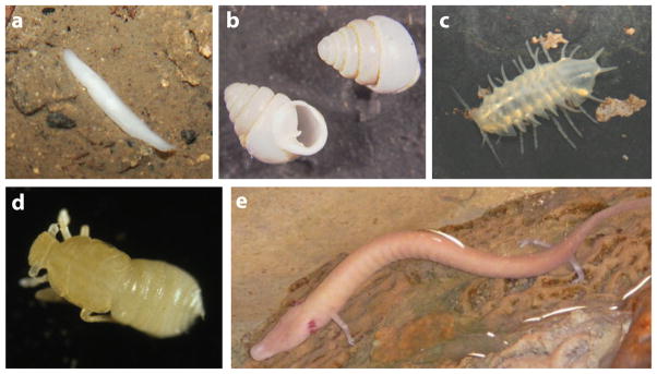
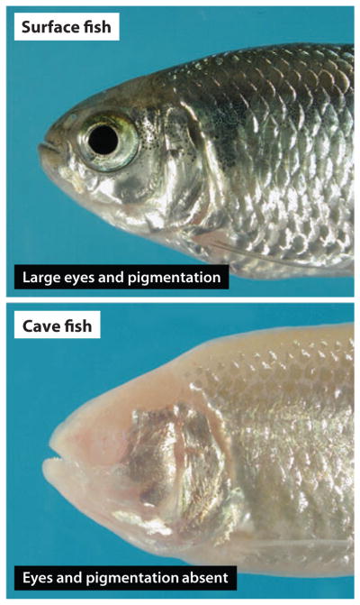
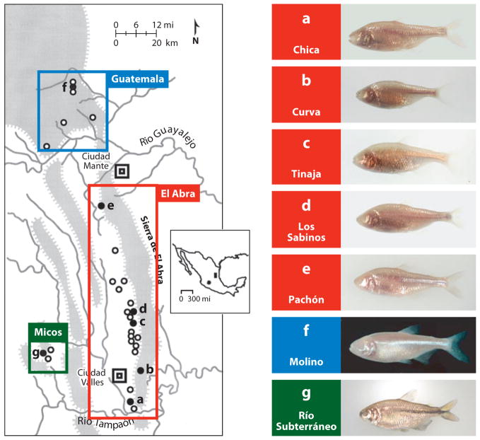
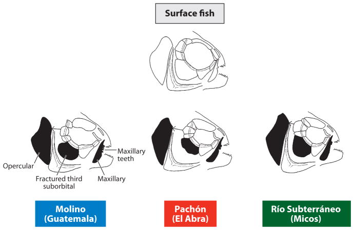
A diverse group of animals, including members of most major phyla, have adapted to life in the perpetual darkness of caves. These animals are united by the convergence of two regressive phenotypes, loss of eyes and pigmentation. The mechanisms of regressive evolution are poorly understood. The teleost Astyanax mexicanus is of special significance in studies of regressive evolution in cave animals. This species includes an ancestral surface dwelling form and many con-specific cave-dwelling forms, some of which have evolved their recessive phenotypes independently. Recent advances in Astyanax development and genetics have provided new information about how eyes and pigment are lost during cavefish evolution; namely, they have revealed some of the molecular and cellular mechanisms involved in trait modification, the number and identity of the underlying genes and mutations, the molecular basis of parallel evolution, and the evolutionary forces driving adaptation to the cave environment.


Figures

Figure 1
Figure 1
Examples of diverse cave animals lacking eyes and pigmentation. (a) Cave planarian. (b) Cave gastropod mollusk, Zospeum kusceri. (c) Cave isopod, Monolistra bolei. (d) Cave planthopper, Oliarus polyphemus. (e) Cave urodele amphibian, Proteus anguinus.
Figure 2
Figure 2
Astyanax mexicanus surface fish and cavefish.
Figure 3
Figure 3
Left: A sketch map showing the region containing 29 different Astyanax cavefish populations in northeastern Mexico. The spheres indicate the approximate position of caves with Astyanax cavefish. The shaded spheres (ag) indicate specific cavefish populations shown on the right. The Guatemala, El Abra, and Micos clusters are indicated on the map. Inset: Mexico showing the northeastern region indicated in the sketch map (shaded rectangle) and the outlying Guerrero population (shaded sphere). Right: Examples of different cavefish shown in ag on the left. Modified from Jeffery et al. (2003).
Figure 4
Figure 4
Surface fish and cavefish exhibit differences in craniofacial morphology. Shaded bones show the most change from surface fish to cavefish and amongst the Guatemala, El Abra, and Micos cavefish clusters. Modified from Yamamoto et al. (2003).
Figure 5
Figure 5
Eye development and degeneration in surface fish and cavefish respectively. (a,b) The lens and optic cup are smaller, and the ventral side of the optic cup is missing in Pachón cavefish embryos (b) relative to surface fish embryos (a). (c,d) TUNEL assay shows apoptosis at 2 days postfertilization (dpf) in the lens and retina of Pachón cavefish embryos (d) but not in surface fish embryos (c). (e, f) PCNA labelling shows active cell proliferation in the CMZ of surface fish (e) and Pachón cavefish (f) retinas at 10 dpf. (g) A diagram showing the events of Astyanax eye development and degeneration. Left. Early events of eye primordium formation are the same in surface fish and cavefish until approximately 1 dpf. Top: In surface fish, the eye differentiates and the eye parts grow in concert with increased body growth. Bottom. In cavefish, the eye primordium grows for a while, then arrests, degenerates, and is internalized by overgrowth of the body. (a,b) from Yamamoto & Jeffery (2000). (c,d) from Strickler et al. (2007). (e, f) from Strickler et al. (2002).
Figure 6
Figure 6
Lens transplantation. (ae) Transplantation of a surface fish embryonic lens into the optic cup of a Pachón (b,c) or Los Sabinos (d,e) cavefish embryo after their own lens is removed rescues the eye in adults. (b,d) Lens transplant side. (c,e) Unoperated side. (fj) Transplantation of a Pachón (g,h) or Los Sabinos (i, j) embryonic lens into the optic cup of a surface fish embryo after its own lens is removed causes retardation of eye development in adults. (g,i) Lens transplant side. (i, j) Unoperated side. From Yamamoto and Jeffery (2000) and Jeffery et al. (2003).
Figure 7
Figure 7
Genetic complementation tests for eye and pigment regression. (a,b) Extent of eye formation in F1 hybrids produced by crosses between different cavefish populations. (a) A Río Subterráneo x Pachón F1 hybrid with large external eyes. (b) Punnett square illustrating eye size in F1 hybrids of crosses between various cavefish populations. (c,d) Extent of pigmentation in F1 hybrids produced by crosses between different cavefish populations. (c) Albinisim is present in a Pachón x Molino F1 hybrid. (d) Punnett square illustrating the status of pigmentation in F1 hybrids of crosses between different cavefish populations. LS: Los Sabinos. Pa: Pachón (albino). Pie: Piedras. Ye: Yerbaniz. Ja: Japonés (albino). Cu: Curva. Mo: Molino (albino). RS: Río Subterráneo. (a) Y. Yamamoto and W.R. Jeffery (unpublished). (c) From Protas et al. (2006).
Figure 8
Figure 8
Negative relationship between midline Shh signaling and Pax6 controls eye development. (a) Diagram of the neural plate and underlying anterior midline of a typical vertebrate embryo showing the negative relationship between Shh (blue) and Pax6 (red). (b,c) Expanded shhA expression along the anterior midline of (c) Pachón cavefish embryo with respect to (b) surface fish embryo hybridized in situ at the neural plate stage. Dorsal views of the anterior neural plate showing shhA expression (blue) compared with dlx3, pax6, and pax2a markers. From Yamamoto et al. (2004).
Figure 9
Figure 9
An anchored linkage map reveals eye and pigment candidate genes in Astyanax QTL and corresponding syntenic regions in Danio (zebrafish). (a) The position of the αA-crystallin gene on Astyanax linkage group 21 and syntenic region on Danio chromosome 1. (b) The positions of the shroom2 and oca2 genes on Astyanax linkage group 4 and syntenic region on Danio chromosome 6. From Gross et al. (2008).
Figure 10
Figure 10
Cavefish tyrosinase-positive albinism. (a) A summary of steps in which L-tyrosine is converted to melanin in the melanosome and dopamine in the cytoplasm showing the key roles of tyrosinase, other enzymes, and the presumed role of OCA2 as an L-tyrosine transporter. (b) Melanophores revealed in an albino Pachón cavefish embryo after conversion of exogenous L-DOPA to melanin by active tyrosinase. TPC: tyrosinase positive cells (melanophores). (c,d) Explants of surface fish (c) albino Pachón adult tail fin (d) showing melanophores in the former but not the latter. (e) Albino Pachón adult tail fin explant in which tyrosinase positive cells are revealed by L-DOPA treatment and melanin deposition. (f) Albino Pachón adult tail fin explant showing inability to convert exogenous L-tyrosine to melanin. The presence of xanthophores is also indicated in albino Pachón adult tail fin explants. (af) from McCauley et al. (2004).
Figure 11
Figure 11
Diagrams indicating the approximate positions (red bars) and types of mutations in genes encoding the OCA2 and Mc1r proteins in different cavefish populations. In OCA2, black regions indicate the 12 membrane spanning domains indicate and blue regions indicate the intervening protein sequences. In Mc1r, black regions indicate the seven membrane spanning domains indicate and pink regions indicate the intervening protein sequences.

Similar articles

Cited by

References

    1. Alunni A, Menuet A, Candal E, Pénigault J-B, Jeffery WR, Rétaux S. Developmental mechanisms for retinal degeneration in the blind cavefish Astyanax mexicanus. J Comp Neurol. 2007;505:221–33. - PubMed
    1. Alvarez J. Revisión del generero Anoptichthys con descriptión de una especies nueva (Pisc. Characidae) An Esc Nac Cien Biol Mexico. 1946;4:263–82.
    1. Alvarez J. Descriptión de Anoptichthys hubbsi caracinído ciego de La Cueva de Los Sabinos, S. L. P. Rev Soc Mexicana Hist Nat. 1947;8:215–19.
    1. Behrens M, Wilkens H, Schmale H. Cloning of the αA-crystallin genes of the blind cave form and the epigean form of Astyanax fasciatus: a comparative analysis of structure, expression and evolutionary conservation. Gene. 1998;216:319–26. - PubMed
    1. Borowsky R. Restoring sight in blind cavefish. Curr Biol. 2008;18:R23–24. - PubMed

Publication types

LinkOut - more resources