

A computational approach identifies two regions of Hepatitis C Virus E1 protein as interacting domains involved in viral fusion process
- PMID: 19640267
- PMCID: PMC2732612
- DOI: 10.1186/1472-6807-9-48
A computational approach identifies two regions of Hepatitis C Virus E1 protein as interacting domains involved in viral fusion process
Abstract
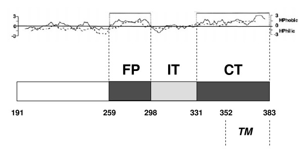
Background: The E1 protein of Hepatitis C Virus (HCV) can be dissected into two distinct hydrophobic regions: a central domain containing an hypothetical fusion peptide (FP), and a C-terminal domain (CT) comprising two segments, a pre-anchor and a trans-membrane (TM) region. In the currently accepted model of the viral fusion process, the FP and the TM regions are considered to be closely juxtaposed in the post-fusion structure and their physical interaction cannot be excluded. In the present study, we took advantage of the natural sequence variability present among HCV strains to test, by purely sequence-based computational tools, the hypothesis that in this virus the fusion process involves the physical interaction of the FP and CT regions of E1.
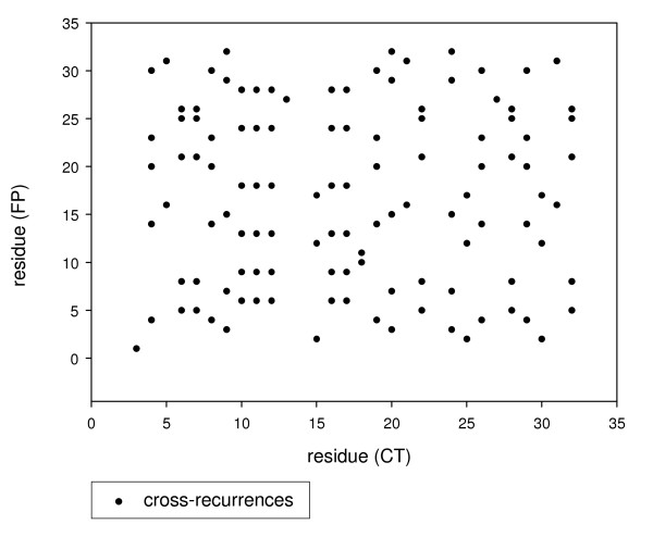
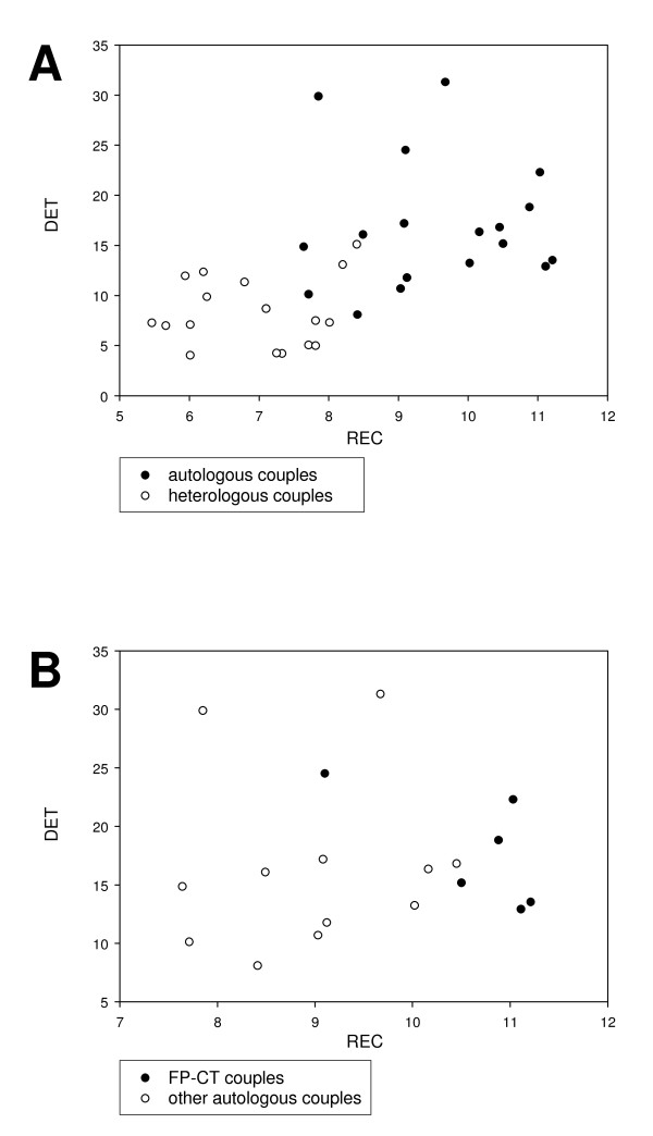
Results: Two computational approaches were applied. The first one is based on the co-evolution paradigm of interacting peptides and consequently on the correlation between the distance matrices generated by the sequence alignment method applied to FP and CT primary structures, respectively. In spite of the relatively low random genetic drift between genotypes, co-evolution analysis of sequences from five HCV genotypes revealed a greater correlation between the FP and CT domains than respect to a control HCV sequence from Core protein, so giving a clear, albeit still inconclusive, support to the physical interaction hypothesis.The second approach relies upon a non-linear signal analysis method widely used in protein science called Recurrence Quantification Analysis (RQA). This method allows for a direct comparison of domains for the presence of common hydrophobicity patterns, on which the physical interaction is based upon. RQA greatly strengthened the reliability of the hypothesis by the scoring of a lot of cross-recurrences between FP and CT peptides hydrophobicity patterning largely outnumbering chance expectations and pointing to putative interaction sites. Intriguingly, mutations in the CT region of E1, reducing the fusion process in vitro, strongly reduced the amount of cross-recurrence further supporting interaction between this region and FP.
Conclusion: Our results support a fusion model for HCV in which the FP and the C-terminal region of E1 are juxtaposed and interact in the post-fusion structure. These findings have general implications for viruses, as any visualization of the post-fusion FP-TM complex has been precluded by the impossibility to obtain crystallised viral fusion proteins containing the trans-membrane region. This limitation gives to sequence based modelling efforts a crucial role in the sketching of a molecular interpretation of the fusion process. Moreover, our data also have a more general relevance for cell biology as the mechanism of intracellular fusion showed remarkable similarities with viral fusion.
Figures
Similar articles
-
Functional Analysis of Hepatitis C Virus (HCV) Envelope Protein E1 Using a trans-Complementation System Reveals a Dual Role of a Putative Fusion Peptide of E1 in both HCV Entry and Morphogenesis.J Virol. 2017 Mar 13;91(7):e02468-16. doi: 10.1128/JVI.02468-16. Print 2017 Apr 1. J Virol. 2017. PMID: 28100619 Free PMC article.
-
Conservation of hydrophobicity within viral envelope glycoproteins reveals a putative hepatitis C virus fusion peptide.Protein Pept Lett. 2009;16(7):815-22. doi: 10.2174/092986609788681779. Protein Pept Lett. 2009. PMID: 19601912
-
Hepatitis C Virus Envelope Glycoprotein E1 Forms Trimers at the Surface of the Virion.J Virol. 2015 Oct;89(20):10333-46. doi: 10.1128/JVI.00991-15. Epub 2015 Aug 5. J Virol. 2015. PMID: 26246575 Free PMC article.
-
Structure and Function of the Hepatitis C Virus Envelope Glycoproteins E1 and E2: Antiviral and Vaccine Targets.ACS Infect Dis. 2016 Nov 11;2(11):749-762. doi: 10.1021/acsinfecdis.6b00110. Epub 2016 Aug 16. ACS Infect Dis. 2016. PMID: 27933781 Review.
-
Peptide entry inhibitors of enveloped viruses: the importance of interfacial hydrophobicity.Biochim Biophys Acta. 2014 Sep;1838(9):2180-97. doi: 10.1016/j.bbamem.2014.04.015. Epub 2014 Apr 26. Biochim Biophys Acta. 2014. PMID: 24780375 Free PMC article. Review.
Cited by
-
Sequence-Based Protein Design: A Review of Using Statistical Models to Characterize Coevolutionary Traits for Developing Hybrid Proteins as Genetic Sensors.Int J Mol Sci. 2024 Jul 30;25(15):8320. doi: 10.3390/ijms25158320. Int J Mol Sci. 2024. PMID: 39125888 Free PMC article. Review.
-
VH1-69 antiviral broadly neutralizing antibodies: genetics, structures, and relevance to rational vaccine design.Curr Opin Virol. 2019 Feb;34:149-159. doi: 10.1016/j.coviro.2019.02.004. Epub 2019 Mar 16. Curr Opin Virol. 2019. PMID: 30884330 Free PMC article. Review.
-
An experimental and computational evolution-based method to study a mode of co-evolution of overlapping open reading frames in the AAV2 viral genome.PLoS One. 2013 Jun 24;8(6):e66211. doi: 10.1371/journal.pone.0066211. Print 2013. PLoS One. 2013. PMID: 23826091 Free PMC article.
-
Capitalizing on knowledge of hepatitis C virus neutralizing epitopes for rational vaccine design.Curr Opin Virol. 2015 Apr;11:148-57. doi: 10.1016/j.coviro.2015.04.001. Epub 2015 Apr 29. Curr Opin Virol. 2015. PMID: 25932568 Free PMC article. Review.
-
Identification and Characteristics of Fusion Peptides Derived From Enveloped Viruses.Front Chem. 2021 Aug 23;9:689006. doi: 10.3389/fchem.2021.689006. eCollection 2021. Front Chem. 2021. PMID: 34497798 Free PMC article. Review.
References
MeSH terms
Substances
LinkOut - more resources
Full Text Sources
