

Clinical evaluation of magnetic resonance imaging in coronary heart disease: the CE-MARC study
- PMID: 19640271
- PMCID: PMC3224948
- DOI: 10.1186/1745-6215-10-62
Clinical evaluation of magnetic resonance imaging in coronary heart disease: the CE-MARC study
Abstract
Background: Several investigations are currently available to establish the diagnosis of coronary heart disease (CHD). Of these, cardiovascular magnetic resonance (CMR) offers the greatest information from a single test, allowing the assessment of myocardial function, perfusion, viability and coronary artery anatomy. However, data from large scale studies that prospectively evaluate the diagnostic accuracy of multi-parametric CMR for the detection of CHD in unselected populations are lacking, and there are few data on the performance of CMR compared with current diagnostic tests, its prognostic value and cost-effectiveness.
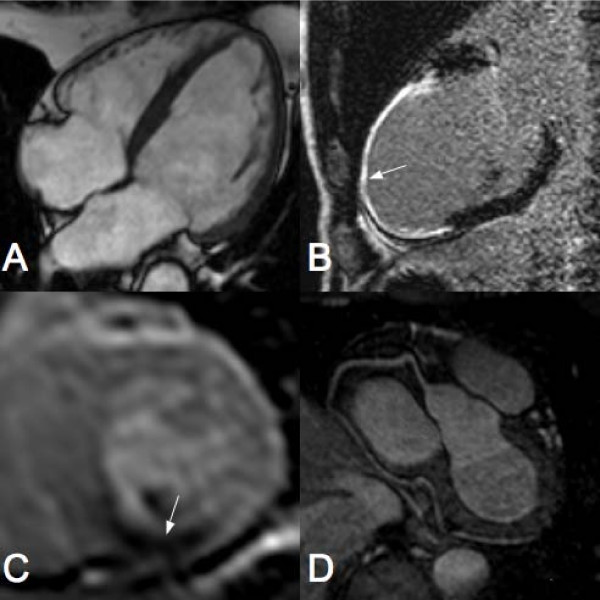
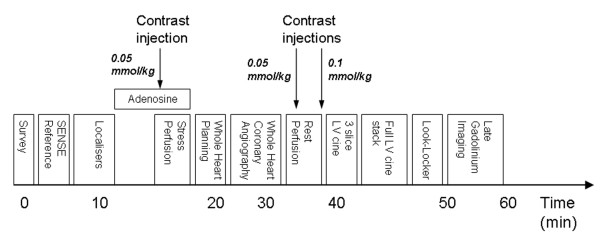
Methods/design: This is a prospective diagnostic accuracy cohort study of 750 patients referred to a cardiologist with suspected CHD. Exercise tolerance testing (ETT) will be preformed if patients are physically able. Recruited patients will then undergo CMR and single photon emission tomography (SPECT) followed in all patients by invasive X-ray coronary angiography. The order of the CMR and SPECT tests will be randomised. The CMR study will comprise rest and adenosine stress perfusion, cine imaging, late gadolinium enhancement and whole-heart MR coronary angiography. SPECT will use a gated stress/rest protocol. The primary objective of the study is to determine the diagnostic accuracy of CMR in detecting significant coronary stenosis, as defined by X-ray coronary angiography. Secondary objectives include an assessment of the prognostic value of CMR imaging, a comparison of its diagnostic accuracy against SPECT and ETT, and an assessment of cost-effectiveness.
Discussion: The CE-MARC study is a prospective, diagnostic accuracy cohort study of 750 patients assessing the performance of a multi-parametric CMR study in detecting CHD using invasive X-ray coronary angiography as the reference standard and comparing it with ETT and SPECT.
Trial registration: Current Controlled Trials ISRCTN77246133.
Figures
References
-
- Allender S, Peto V, Scarborough P, Boxer A, Rayner M. Coronary heart disease statistics. British Heart Foundation Health Promotion Research Group and Department of Public Health, University of Oxford; 2007.
-
- Gianrossi R, Detrano R, Mulvihill D, Lehmann K, Dubach P, Colombo A, McArthur D, Froelicher V. Exercise-induced ST depression in the diagnosis of coronary artery disease. A meta-analysis. Circulation. 1989;80:87–98. - PubMed
-
- Myocardial perfusion scintigraphy for the diagnosis and management of angina and myocardial infarction. National Institute for Clinical Excellence; 2003.
Publication types
MeSH terms
Associated data
Grants and funding
LinkOut - more resources
Full Text Sources
Medical
