. 2009 Sep 15;392(1):34-51.
doi: 10.1016/j.virol.2009.07.002. Epub 2009 Jul 28.

Kaposi sarcoma-associated herpes virus (KSHV) G protein-coupled receptor (vGPCR) activates the ORF50 lytic switch promoter: a potential positive feedback loop for sustained ORF50 gene expression

Affiliations

Kaposi sarcoma-associated herpes virus (KSHV) G protein-coupled receptor (vGPCR) activates the ORF50 lytic switch promoter: a potential positive feedback loop for sustained ORF50 gene expression

Virginie Bottero et al. Virology. .

Abstract

KSHV vGPCR, a lytic cycle associated protein, induces several signaling pathways leading to the activation of various transcription factors and consequently the expression of cellular and viral genes. Though the role of vGPCR in KSHV tumorigenicity has been well studied, its function related to the viral life cycle is poorly understood. Reduction in vGPCR by RNA interference also resulted in the reduction in KSHV lytic switch ORF50 gene and protein expression. Induction of vGPCR by doxycycline in BC3.14 cells also resulted in more KSHV production. When this was explored, induction of the ORF50 promoter by vGPCR expression was observed. Further examination of the molecular mechanisms by which vGPCR regulates the ORF50 promoter, using various ORF50 promoter constructs, revealed that induction of ORF50 promoter by vGPCR did not involve AP1 but was dependent on Sp1 and Sp3 transcription factors. vGPCR signaling led to an increase in Sp1 and Sp3 DNA binding activity and a decrease in histone deacetylase (HDAC) activity. These activities were pertussis toxin independent, did not involve Rho and Rac-GTPases and involved the heterotrimeric G protein subunits Galpha12 and Galphaq. Studies using pharmacologic inhibitors and dominant-negative proteins identified phospholipase C, the novel protein kinase C (novel PKC) family and protein kinase D (PKD) as part of the signaling initiated by vGPCR leading to ORF50 promoter activation. Taken together, this study suggests a role for vGPCR in the sustained expression of ORF50 which could lead to a continued activation of lytic cycle genes and ultimately to successful viral progeny formation.


Figures
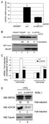
Figure 1
Figure 1
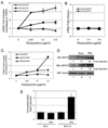
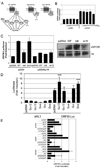
vGPCR silencing. (A) HEK293T cells were transfected with vGPCR expressing vector together with a plasmid expressing an siRNA control against luciferase (si-C) or siRNA against vGPCR (si-vGPCR-1). RNAs were prepared, reverse transcribed into cDNA, levels of vGPCR measured by quantitative real-time RT-PCR and normalized to the levels of the house keeping gene tubulin. (B) HEK293T cells were transduced with a lentivector expressing a control siRNA (si-C) or a siRNA against vGPCR (si-vGPCR-1). The lentivector also expressed GFP under a CMV promoter thus allowing for measurement of the level of transduction. One week later, the cells were transfected with an empty expression plasmid or a plasmid expressing vGPCR. 48h later, the cells were lysed in RIPA lysis buffer and 50 µg of protein were subjected to Western blot analysis. Detection of tubulin was used as a loading control and GFP as transduction control. (C) BCBL-1 cells were transduced with the lentivector expressing the control siRNA against luciferase (si-C) or with the lentivector expressing the siRNA against vGPCR (si-vGPCR). Both lentivectors also expressed GFP under a constitutive promoter allowing for the estimation of the level of transduction by fluorescence microscopy and flow cytometry. One week after transduction, the cells were treated with TPA (20 ng/ml) for 0 or 4 days. The cells were then assayed for accumulation of vGPCR and ORF50 mRNA by quantitative real-time RT-PCR. Each reaction was done in triplicate and a representative experiment of three experiments is shown here. +/− SD; ***p<0.0005. (D) BCBL-1 cells were transduced with the lentivector expressing si-C or si-vGPCR. After one week, cells were untreated or treated with TPA (20 ng/ml) for 3 days. Total protein extracts were prepared and the level of ORF50 and vGPCR expression was assessed by western blot. Tubulin was used as loading control.
Figure 2
Figure 2
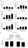
Induction of vGPCR and KSHV lytic gene expression. (A) BC3.14 cells were treated for 48h with the indicated doses of doxycycline (0, 0.05, 0.1 and 0.5 µg/ml). The cells were then assayed for accumulation of vGPCR, ORF50, and ORF73 mRNAs by quantitative real-time RT-PCR. Results were normalized to HPRT. Each reaction was done in triplicate and the data represent the mean of four independent experiments +/− S.E.M. (B) BC3 cells were treated for 48h with the indicated doses of doxycycline (0, 0.1 and 0.5 µg/ml) and assayed for the accumulation of vGPCR and ORF50 mRNAs by quantitative real-time RT-PCR. Results were normalized to HPRT. Each reaction was done in triplicate and the data represent the mean of three independent experiments +/− S.E.M. (C) BC3.14 cells were treated for 48h with the indicated doses of doxycycline (0, 0.05, 0.1 and 0.5 µg/ml). The cells were then assayed for the accumulation of PAN, ORF57, and ORF26 mRNAs by quantitative real-time RT-PCR. Results were normalized to HPRT. Each reaction was done in triplicate and the data represent the mean of three independent experiments +/−S.E.M. (D) BC3.14 cells were treated for 3 days with TPA (20 ng/ml) or doxycycline (1 µg/ml). Total proteins were extracted in RIPA lysis buffer and subjected to a western blot experiment using the indicated antibodies. (E) BC3 and BC3.14 cells were treated for 2 days with doxycycline (0.5 µg/ml). After centrifugation for 10 minutes at 2000 rpm, the supernatant was filtered on a 0.45 µm filter. Virus quantification was performed by real time PCR quantitation of extracted viral DNA (ORF73 gene).
Figure 3
Figure 3
(A–F) Effect of vGPCR on different KSHV promoters. HEK293T cells were transfected by the calcium phosphate method with pcDNA or an increasing quantity of vGPCR expression plasmids (0, 0.5, 1 or 2 µg), and 1 µg of the indicated promoter luciferase constructs (A: pGL3, B: ORF50-Luc; C: PAN-Luc; D: LanaPc-Luc; E: LanaPi-Luc; F: K14-Luc), and 0.5 µg of the β-gal reporter construct (as transfection control). Cells were harvested 36h after transfection. The luciferase and β-galactosidase activities were measured in triplicate using Steady-glo and Beta-glo Promega kits, respectively. The luciferase activity was normalized to β-galactosidase activity as transfection control. The data represent the mean of fold induction of at least three independent experiments +/− S.E.M. ** p<0.05. ***p<0.01. (G) HEK293T cell were transfected by the calcium phosphate method with 3 µg of pcDNA or vGPCR expression plasmids. The same day, other HEK293T cells were transfected with 2 µg of the ORF50 promoter luciferase constructs and 1 µg of the β-Gal reporter construct as transfection control. The day after transfection, the medium was replaced by fresh medium and was collected 24h later. The medium isolated from pcDNA or vGPCR transfected cells was added to the cells transfected with the reporter constructs. 8h or 16h later, the luciferase and β-galactosidase activities were measured in triplicate using Steady-glo and Beta-glo Promega kits, respectively, and the luciferase activity was normalized to β-galactosidase activity. The data represent the mean of fold induction of five independent experiments +/− S.E.M.
Figure 4
Figure 4
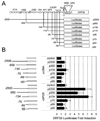
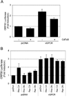
(A and B) Mapping of ORF50 promoter elements involved in vGPCR induced activation. HEK293T cells were transfected by the calcium phosphate method with 2 µg of pcDNA or vGPCR expression plasmids, 1 µg of the indicated ORF50 promoter luciferase constructs, and 0.5 µg of the β-gal reporter construct. Cells were harvested 36h after transfection, the luciferase and β-galactosidase activities measured in triplicate, and the luciferase activity was normalized to β-galactosidase activity. The data represent the mean of fold induction of four independent experiments +/− S.E.M.
Figure 5
Figure 5
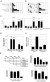
Sp1, but not AP1, activates the ORF50 promoter after vGPCR expression. (A and B) HEK293T cells were transfected by the calcium phosphate method with 2 µg of pcDNA or vGPCR expression plasmids, 1 µg of the indicated ORF50 promoter luciferase constructs (A: ORF50 promoters with deleted AP1 or Sp1 sites; B: ORF50 promoters with mutated AP1 or Sp1 sites), and 0.5 µg of the β-gal reporter construct. Cells were harvested 36h after transfection. The luciferase and β-galactosidase activities were measured in triplicate and the luciferase activity was normalized to β-galactosidase activity. The data represent the mean of fold induction of three independent experiments +/− S.E.M. (C) Effect of vGPCR induction on Sp1 and Sp3 transcription factor activation. Nuclear extracts prepared from untreated or doxycycline treated BC3.14 cells were tested for the activation of Sp1 and Sp3 transcription factors by incubating the nuclear extracts with plate-immobilized oligonucleotides containing the Sp1 family consensus binding sequence, which was followed by ELISA with antibodies to the respective transcription factors. MCF7 (human breast adenocarcinoma cells) nuclear extracts provided in the Sp1/3 ELISA kit were used as a positive control. To confirm the specificity of binding, the nuclear extracts were incubated with WT or mutated oligonucleotide prior to the plate-immobilized oligonucleotide incubation. The data represent the average and standard deviations of two independent experiments. *** p<0.001. (D) HEK293T cells were transfected by the calcium phosphate method with 1 µg of the ORF50 promoter luciferase construct WT (p134) or mutated at the Sp1/3 site (p134Sp1M), and 0.5 µg β-galactosidase reporter, in the presence or absence of 2 µg of Sp1 or Sp3 expression plasmids. 36h after transfection, luciferase and β-galactosidase were measured in triplicate and the data represent the mean fold induction of four independent experiments +/− S.E.M. ** p<0.01. *** p<0.0005. (E) HEK293T cells were transfected by the calcium phosphate method with 1 µg of the ORF50 promoter luciferase construct WT (p2500) or mutated at the Sp1/3 site (p2500Sp1M), and 0.5 µg β-galactosidase reporter, in the presence or absence of 2 µg of Sp1 expression plasmids. 36h after transfection, luciferase and β-galactosidase were measured in triplicate and the data represented as the mean fold induction of three independent experiments +/− S.E.M. (F) Mapping of PAN promoter elements involved in vGPCR-induced activation. HEK293T cells were transfected by the calcium phosphate method with 2 µg of pcDNA or vGPCR expression plasmids, 1 µg of the indicated PAN promoter luciferase constructs, and 0.5 µg of β-gal reporter construct. Cells were harvested 36h after transfection and the luciferase and β-galactosidase activities measured in triplicate. Luciferase activity was normalized to β-galactosidase activity. The data represent the mean of fold induction of four independent experiments +/− S.E.M.
Figure 6
Figure 6
Regulation of the ORF50 promoter after vGPCR expression is partially mediated by Gαq and Gα12. (A) Schematics depicting wild-type and mutant vGPCR (m15 linked to Gαq and m8 linked to Gαi) (B) ORF50 regulation by vGPCR expression is pertussis toxin (PTx) insensitive: HEK293T cells were transfected by the calcium phosphate method with 2 µg of pcDNA or vGPCR expression plasmids along with 1 µg of full length ORF50-luciferase reporter (p2500Luc) and 0.5 µg of the β-gal reporter plasmid (as transfection control). 24h post-transfection, the cells were treated overnight with increasing doses of PTx (0, 50, 100, 200, and 1000 ng/ml). The data represent the mean of fold induction of three independent experiments +/− S.E.M. (C) Wild-type and mutant vGPCR (m15, linked to Gαq) can activate the ORF50 promoter. HEK293T cells were transfected by the calcium phosphate method with 2 µg of pcDNA or the indicated vGPCR expression plasmids (WT, m8, m15) along with 1µg of the full length ORF50-luciferase reporter (p2500) or its Sp1 mutated version (p2500Sp1M) and 0.5 µg of the β-gal reporter plasmid (as transfection control). 36h after transfection, luciferase and β-galactosidase were measured in triplicate and the data represent the mean of fold induction of three independent experiments +/− S.E.M. In addition, total proteins were extracted in RIPA lysis buffer and subjected to a western blot experiment using vGPCR antibodies (right panel). (D) Expression of constitutively active mutants Gα12 and Gαq activate the ORF50 promoter. HEK293T cells were transfected by the calcium phosphate method with 1 µg of the indicated luciferase reporter (pGL3 or ORF50 full length promoter p2500Luc), 0.5 µg of the β-galactosidase reporter (as transfection control) and 2 µg of the indicated G protein expression plasmids. 36h after transfection, luciferase and β-galactosidase were measured in triplicate and the data represent the mean of fold induction of five independent experiments +/− S.E.M. *** p<0.005. (E) The Sp1 site is involved in Gα12 and Gαq activation of the ORF50 promoter. HEK293T cells were transfected by the calcium phosphate method with 2 µg of pcDNA or Gα12QM or GαqQM expression plasmids, 1 µg of the indicated ORF50 promoter luciferase constructs, and 0.5 µg of the β-Gal reporter construct (as transfection control). Cells were harvested 36h after transfection. The luciferase and β-galactosidase activities were measured in triplicate and the luciferase activity was normalized to β-galactosidase activity. The data represent the mean of fold induction of three independent experiments +/− S.E.M.
Figure 7
Figure 7
G-proteins Rho and Rac are not involved in ORF50 promoter regulation after vGPCR expression. (A) HEK293T cells were transfected by the calcium phosphate method with 1 µg of ORF50 full length promoter p2500Luc, 0.5 µg of the β-galactosidase reporter (as transfection control) in the presence or absence of 2 µg of the vGPCR expression construct. 24h after transfection, the cells were incubated with or without CdTxB (200 ng/ml) for 16h. Luciferase and β-galactosidase were measured in triplicate and the data represent the mean of fold induction of three independent experiments +/− S.E.M. (B) HEK293T cells were transfected by the calcium phosphate method with 1 µg of the ORF50 full length promoter p2500Luc, 0.5 µg β-galactosidase reporter (as transfection control) and 1 µg of the indicated expression plasmids, in the presence or absence of 1 µg of the vGPCR expression construct. 36h after transfection, luciferase and β-galactosidase were measured in triplicate and the data represent the mean fold induction of four independent experiments +/− S.E.M.
Figure 8
Figure 8
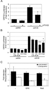
Effect of pharmacologic signaling pathway inhibitors. (A) Effect of PLC inhibitor. HEK293T cells were transfected by the calcium phosphate method with 1 µg of the ORF50 full length promoter p2500Luc, 0.5 µg of the β-galactosidase reporter in the presence or absence of 2 µg of the vGPCR expression construct. 24h after transfection, the cells were incubated with or without the indicated PLC inhibitor (U73122; 10 µM) for 16h. Luciferase and β-galactosidase were measured in triplicate and the data represent the mean of fold induction of four independent experiments +/− S.E.M. p<0.05. (B) Effect of PKC inhibitors. HEK293T cells were transfected by the calcium phosphate method with 1 µg of the ORF50 full length promoter p2500Luc, 0.5 µg of the β-galactosidase reporter in the presence or absence of 2 µg of the vGPCR expression construct. 24h after transfection, the cells were incubated with or without the indicated PKC inhibitors (Rottlerin 10 µM, GFX 10 µM, Gö6983 1 or 10 µM) for 16h. Luciferase and β-galactosidase were measured in triplicate and the data represent the mean of fold induction of three independent experiments (Gö6983) or 5 experiments (GFX and Rottlerin) +/− S.E.M. * p<0.05, ***p<0.0005. (C) BC3.14 cells were treated for 48h with or without doxycycline (0.5 µg/ml) in presence or absence of the indicated PKC inhibitors (Rottlerin 10 µM, GFX 10 µM). The cells were then assayed for accumulation of vGPCR and ORF50 mRNAs by quantitative real-time RT-PCR. Results were normalized to HPRT and the fold induction with / without doxycycline is presented. Each reaction was done in triplicate and the data represent the mean of three independent experiments.
Figure 9
Figure 9
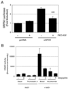
Effect of PKD and HDAC. (A) HEK293T cells were transfected by the calcium phosphate method with 1 µg of the ORF50 full length promoter p2500Luc, and 0.5 µg β-galactosidase reporter in the presence or absence of 1 µg of PKD-KW expression plasmids as well as in the presence or absence of 1 µg of the vGPCR expression construct. 36h after transfection, luciferase and β-galactosidase were measured in triplicate and the data represent the mean fold induction of four independent experiments +/− S.E.M. ** p<0.01. (B) BC3.14 cells were treated with Doxycycline (0.5 µg/ml) for 48h. 1 µg of nuclear extracts were used in HDAC assays as described in the Material and Methods section. Briefly, nuclear extracts were incubated in the absence or presence of 250 µM NAD+ cofactor. After HDAC deacetylation of the substrate, a fluorophore is released and detected using a fluorescence plate reader (Synergy™ HT Multi-Mode Microplate Reader). Specificity of HDAC activity was checked using the HDAC inhibitor trichostatin A (1 µM). Specificity of the class III HDAC activity was analyzed in the presence of metabolite nicotinamide (5 µM). The data represent the mean fluorescent intensity of two independent experiments with the fluorescence read in triplicate ± S.D.
Figure 10
Figure 10
Schematic representation depicting the mechanism of ORF50 promoter regulation induced by vGPCR. The constitutive activity of vGPCR through the Gαq and Gα12 proteins leads to the activation of the PLC and PKC signaling pathways, which directly increase the nuclear level of Sp1 and Sp3 transcription factors as well as their binding to their specific target sites present on the ORF50 promoter. In addition, vGPCR releases the ORF50 promoter repression induced by HDAC and the inhibition of HDAC could involve a phosphorylation step induced by PKD. Together, these activities of vGPCR could act as part of a positive feedback loop resulting in the sustained expression of ORF50, which could lead to continued activation of ORF50 dependent lytic cycle genes and to successful viral progeny formation.

Similar articles

Cited by

References

    1. Aktories K. Bacterial toxins that target Rho proteins. J Clin Invest. 1997;99(5):827–829. - PMC - PubMed
    1. Alberts BLJ, Raff M, Roberts K, Walter P. Molecular biology of the cell. 2002
    1. Bais C, Van Geelen A, Eroles P, Mutlu A, Chiozzini C, Dias S, Silverstein RL, Rafii S, Mesri EA. Kaposi's sarcoma associated herpesvirus G protein-coupled receptor immortalizes human endothelial cells by activation of the VEGF receptor-2/ KDR. Cancer Cell. 2003;3(2):131–143. - PubMed
    1. Bhende PM, Dickerson SJ, Sun X, Feng WH, Kenney SC. X-box-binding protein 1 activates lytic Epstein-Barr virus gene expression in combination with protein kinase D. J. Virol. 2007;81(14):7363–7370. - PMC - PubMed
    1. Borras AM, Strominger JL, Speck SH. Characterization of the ZI domains in the Epstein-Barr virus BZLF1 gene promoter: role in phorbol ester induction. J Virol. 1996;70(6):3894–3901. - PMC - PubMed

Publication types

MeSH terms